Tag Archives: angiography

Spleen Injury, Angiography, And Splenectomy

The shift toward initial nonoperative management of spleen injuries began in the early 1990’s, as the resolution of early CT scans began to improve. Our understanding of the indicators of failure also improved over time, and success rates rose and splenectomy rates fell.

Angiography was adopted as an adjunct to early management, especially when we figured out what contrast extravasation and pseudoaneurysms really meant (bad news, and nearly certain failure in adults). At first, it was used in a shotgun approach in most of the higher grade injuries. But we have refined it over the years, and now it is used far more selectively at most centers.

A group at Indiana University was interested in looking at the impact of angio use on splenic salvage over a long time frame. They queried the National Trauma Data Bank, looking specifically at high grade splenic injury care at Level I and II centers from 2008-2014. Patients undergoing splenectomy were divided into early (<= 6hr after admission) and late (> 6 hrs). Over 50,000 records were analyzed.

Here are the factoids:

  • There was a shift from early splenectomy to late splenectomy over the study period that was statistically significant
  • Use of angio increased from 5 to 12% during the study period
  • Overall splenectomy rate remained about the same

So the authors recognize that late splenectomy has decreased. But they also state that early splenectomy has increased. They attribute it to increased recognition of patient requiring early splenectomy. They then call into question the need to use angiography if it hasn’t decreased the overall splenectomy rate.

Problem: The early splenectomy rate increased from about 13% to 14%, reading their graph, and is probably not significant. These are the failures that occur in the trauma bay and shortly thereafter that must be taken to the OR. The late splenectomy rate decreased from 5% to 3%, which may be significant (p value not included in the abstract). These are failures during nonoperative management, and are decreasing over time. And BTW, the authors do not define what “high grade” splenic injuries they are looking at.
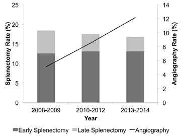
AAST2016-Paper35

Bottom line: This abstract illustrates why it is important to read the entire article, or in this case, listen to the full presentation at AAST. It sounds like one that’s been written to justify not having angiography available as it is currently required. 

The authors showed that overall splenectomy rate was the same, but delayed splenectomy (late failure) has decreased with increasing use of angiography. But remember, this is an association, not cause and effect. Most of the early failures are still probably ones that can’t be prevented, but we’ll see if the authors can dissect out how many went to OR very early (not eligible for angio), or later in the 6 hour period (could have used angio). It looks to me like the use of angiography is having the desired effect. But undoubtedly we could use that resource more wisely. What we really need are some guidelines as to exactly when a call to the interventional radiologists is warranted.

Related posts:

Reference: Overall splenectomy rates remain the same despite increasing usage of angiography in the management of high grade blunt splenic injury. AAST 2016, paper 35.

IV Contrast In Trauma Imaging

We use CT scanning in trauma care so much that we tend to take it (and its safety) for granted. I’ve written quite a bit about thoughtful use of radiographic studies to achieve a reasonable patient exposure to xrays. But another thing to think about is the use of IV contrast.

IV contrast is a hyperosmolar solution that contains some substance (usually an iodine compound) that is radiopaque to some degree. It has been shown to have a significant impact on short-term kidney function and in some cases can cause renal failure.

Here are some facts you need to know:

  • Contrast nephrotoxicity is defined as a 25% increase in serum creatinine, usually within the first 3 days after administration
  • There is usually normal urine output and minimal to no proteinuria
  • In most cases, renal function returns to normal after 3-4 days
  • Nephrotoxicity almost never occurs in people with normal baseline kidney function
  • Large or repeated doses given within 72 hours greatly increase risk for toxicity
  • Old age and pre-existing diabetic renal impairment also greatly increase risk

If you must give contrast to a patient who is at risk, make sure they are volume expanded (tough in trauma patients), or consider giving acetylcysteine or using isosmolar contrast (controversial, may still cause toxicity).

Bottom line: If you are considering contrast CT, try to get a history to see if the patient is at risk for nephrotoxicity. Also consider all of the studies that will be needed and try to consolidate your contrast dosing. For example, you can get CT chest/abdomen/pelvis and CT angio of the neck with one contrast bolus. Consider low dose contrast injection if the patient needs formal angiographic studies in the IR suite. Always think about the global needs of your patient and plan accordingly (and safely).

Related posts:

Reference: Contrast media and the kidney. British J Radiol 76:513-518, 2003.

Unstable Patient & Pelvic Fracture + Hemoperitoneum

The usual thinking is that most unstable trauma patients need a quick trip to the OR to stop the bleeding from something. In the US and Europe, patients with nasty pelvic fractures are no exception, especially those with hemoperitoneum. But many of these patients are bleeding from vessels associated with the pelvic fractures and not so much from associated intra-abdominal injuries. And operative management of pelvic fracture bleeding is far from satisfying, even when using preperitoneal packing.

Well, things are a little different in Japan. In many cases, unstable patients are taken to interventional radiology for angio and possible embolization. Is this prudent, or is it dangerous? A Japanese group decided to critically look at this practice by examining the Japan Trauma Data Bank for answers.

Here are the factoids:

  • Patients with pelvic fracture and positive FAST were included, who underwent either laparotomy or angioembolization as their first intervention (n=1153). Those with non-salvageable head injury were excluded, as well as patients who underwent another major procedure first (craniotomy, thoracotomy, ortho procedures, etc.). Only 317 patients remained.
  • In-hospital mortality was the primary outcome of interest
  • A total of 123 underwent laparotomy first, and 194 went to angio first
  • A very small number of patients were hypotensive on arrival (81 laparotomy first, 82 angio first)
  • Half of the patients who were hypotensive on arrival went to angio first (!)
  • Laparotomy-first patients had a higher crude mortality, but this disappeared when confounders were controlled. This was true in patients who were either normotensive or hypotensive on arrival.
  • The authors concluded that the initial intervention should be determined by severity of injury, since in-hospital mortality was no different

Bottom line: Whoa! This is a sweeping statement for a study with so few subjects. Yes, it can be very difficult to determine whether initial bleeding is from the pelvis vs a solid organ or mesenteric injury while in the ED. But it is all too easy to fritter away time (and the patient’s blood/life) in the angiography suite. I recommend trying to stabilize your patient as best you can with fluid and/or blood. If you can maintain a “reasonable” blood pressure, proceed to CT for a quick look at the torso. Then go to the most appropriate location to take care of the problem. And if your patient decompensates in CT or angio, immediately proceed to the operating room!

Related posts:

References:

  • Comparison between laparotomy first versus angiographic embolization first in patients with pelvic fracture and hemoperitoneum: a nationwide observational study from the Japan Trauma Data Bank. Scand J Trauma 21:82, 2013.
  • Eastern Association for the Surgery of Trauma Practice Management Guidelines for Hemorrhage in Pelvic Fracture-Update and Systematic Review. J Trauma 71:1850-1868, 2011.

Contrast Blush in Children

A contrast blush is occasionally seen on abdominal CT in patients with solid organ injury. This represents active arterial extravasation from the injured organ. In most institutions, this is grounds for call interventional radiology to evaluate and possibly embolize the problem. The image below shows a typical blush from extravasation.

Splenic contrast blush

This thinking is fairly routine and supported by the literature in adults. However, it cannot be generalized to children!

Children have more elastic tissue in their spleen and tend to do better with nonoperative management than adults. The same holds true for contrast blushes. The vast majority of children will stop bleeding on their own, despite the appearance of a large blush. In fact, if children are taken to angiography, it is commonplace for no extravasation to be seen!

Angiography introduces the risk of local complications in the femoral artery as well as more proximal ones. That, coupled with the fact that embolization is rarely needed, should keep any prudent trauma surgeon from ordering the test. It should be reserved for cases where nonoperative management is failing, but hypotension (hard fail) has not yet occurred.

The only difficult questions is “when is a child no longer a child?” Is there an age cutoff at which the spleen starts acting like an adult and keeps on bleeding? Unfortunately, we don’t know. I recommend that you use the “eyeball test”, and reserve angiography for kids with contrast extravasation who look like adults (size and body habitus).

Reference: What is the significance of contrast “blush” in pediatric blunt splenic trauma? Davies et al. J Pediatric Surg 2010 May; 45(5):916-20.

Best Of: IV Contrast

We use CT scanning in trauma care so much that we tend to take it (and its safety) for granted. I’ve written quite a bit about thoughtful use of radiographic studies to achieve a reasonable patient exposure to xrays. But another thing to think about is the use of IV contrast.

IV contrast is a hyperosmolar solution that contains some substance (usually an iodine compound) that is radiopaque to some degree. It has been shown to have a significant impact on short-term kidney function and in some cases can cause renal failure.

Here are some facts you need to know:

  • Contrast nephrotoxicity is defined as a 25% increase in serum creatinine, usually within the first 3 days after administration
  • There is usually normal urine output and minimal to no proteinuria
  • In most cases, renal function returns to normal after 3-4 days
  • Nephrotoxicity almost never occurs in people with normal baseline kidney function
  • Large or repeated doses given within 72 hours greatly increase risk for toxicity
  • Old age and pre-existing diabetic renal impairment also greatly increase risk

If you must give contrast to a patient who is at risk, make sure they are volume expanded (tough in trauma patients), or consider giving acetylcysteine or using isosmolar contrast (controversial, may still cause toxicity).

Bottom line: If you are considering contrast CT, try to get a history to see if the patient is at risk for nephrotoxicity. Also consider all of the studies that will be needed and try to consolidate your contrast dosing. For example, you can get CT chest/abdomen/pelvis and CT angio of the neck with one contrast bolus. Consider low dose contrast injection if the patient needs formal angiographic studies in the IR suite. Always think about the global needs of your patient and plan accordingly (and safely).

Related posts:

Reference: Contrast media and the kidney. British J Radiol 76:513-518, 2003.