Category Archives: Pelvis

Preperitoneal Packing vs Angioembolization For Pelvic Fracture

In my last post, I laid out the various options available for initial management of major pelvic fracture bleeding. Today, I’ll compare two of the newer tools: embolization (AE) and preperitoneal packing (PPP). In the next post, I’ll look at the data available for REBOA.

Interestingly, the use of AE and PPP vary geographically. Angioembolization has been a mainstay in the US for some time, and PPP has been more commonly used in Europe. The use of both is becoming more widespread, and each has its pros and cons.

Angioembolization requires the presence of a special interventional radiology team and a reasonably stable patient. The procedure can take some time, and the IR suite is not really the place to house an unstable patient. Preperitoneal packing requires a reasonably stable pelvis to hold the packs in place for optimal tamponade, which may require application of an external fixator at the time of the procedure.

But is one better than the other? A number of relatively small studies have been performed, which means that it is time to synthesize them and see if some clearer answers can be found. The trauma group in Newcastle, Australia did just this. They performed a systematic search of the literature, analyzing the impact of each procedure on in-hospital mortality.

Here are the factoids:

  • A total of 18 studies met the authors’ inclusion criteria: 6 studies on AE, 9 studies of PPP, and 3 that compared them to each other
  • ISS was significantly higher in the PPP group vs AE (41 vs 36)
  • Average time to OR in the PPP patients was 60 minutes vs 131 minutes to IR in the AE group (statistically significant)
  • A quarter (27%) of the PPP patients did not get adequate hemorrhage control and required AE
  • In-hospital mortality in the PPP papers was 23% vs 32% in the AE research
  • Mortality in the papers that compared AE directly to PPP wasno different

Bottom line: What does this all mean? Is packing “better” than embolization? The simple answer is that we don’t know yet. Due to the way this study was performed, it is not possible to tease out all of the possible confounders. 

We are taught that control of hemorrhage is paramount. The time to definitive management in the AE group was twice that of the PPP patients. This could have a major impact on mortality. Two hours of bleeding can certainly kill. And the lower mortality in the PPP group occurred even though their injury severity was higher.

Many trauma centers have both of these interventions available to choose from. How should we approach their use? Unfortunately the literature is still to scarce to come to strong conclusions. Until we have better research to learn from, I suggest the following:

  • Time is of the essence. Which procedure can you get the fastest? In many cases, this will be preperitoneal packing since it’s just a trip to your trauma OR, which should be ready and waiting. If you have an IR team standing by or available very quickly, you could consider them first.
  • Pay attention to hemodynamic stability. An IR suite is no place for an unstable patient. The resuscitation equipment is not on par with the OR, and one never knows exactly how long the procedure will last.
  • If you have a hybrid room, use it! This is the ideal situation. The surgeon can start the PPP while the orthopedic surgeon applies a fixator. And the radiologist can be preparing to finish it off with a quick squirt as soon as they move away from the groin.
  • The use of one does not rule out the other. If one fails and the patient has increasing fluid and blood requirements move immediately to the other procedure to try to get control.

Reference: Preperitoneal packing versus Angioembolization for the initial management of hemodynamically unstable pelvic fracture – A Systematic Review and Meta-Analysis. J Trauma, publish ahead of print, Jan 4 2022, doi: 10.1097/TA.0000000000003528.


Print Friendly, PDF & Email

The Peri-Mortem C-Section

The perimortem C-section (PMCS) is a heroic procedure designed to salvage a viable fetus from a moribund mother. Interestingly, in some mothers, delivery of the fetus results in return of spontaneous circulation.

The traditional teaching is that PMCS should be started within 4-5 minutes of the mother’s circulatory arrest. The longer it is delayed, the (much) lower the likelihood that the fetus will survive.

The reality is that it takes several minutes to prepare for this procedure because it is done so infrequently in most trauma centers. Recent literature suggests the following management for pregnant patients in blunt traumatic arrest (BTA):

  • Cover the usual BTA bases, including securing the airway, obtaining access and rapidly infusing crystalloid, decompressing both sides of the chest, and assessing for an unstable pelvis
  • Assess for fetal viability. The fundus must measure at least 23 cm.
  • Assess for a shockable vs non-shockable rhythm. If shockable, do two cycles of CPR before beginning the PMCS. If non-shockable, move straight to this procedure.

Bottom line: Any time you receive a pregnant patient in blunt arrest, have someone open the C-section pack while you assess and try to improve the mother’s viability. As soon as you complete the three tasks above, start the procedure! You don’t need to wait 4 minutes! And by the way, this is usually a procedure for surgeons only. They have the speed and skills to get to the right organs quickly. If unavailable, do what you need to do but recognize that the outcome may be even worse than it usually is.

Print Friendly, PDF & Email

Early Operative Fixation of Pelvic Fractures And Functional Outcome

Disruption of the pelvic bones takes a huge amount of energy, and results in significant bleeding and morbidity from other causes. Repair typically consists of surgical fixation, frequently with temporary external fixation in the interim. These patients require intensive therapy postoperatively, with inpatient rehab prior to discharge home.

How well do patients with severe pelvic fractures do in the longer term? The group at the University of Tennessee in Memphis did a lengthy followup study spanning 18 years of severe pelvic fractures treated at their hospital. These patients had sustained fractures with significant bleeding, an open book component, or SI joint disruption with vertical shear.

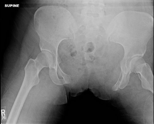
open book pelvis pre

The authors used phone interviews and a standardized measurement instrument (Activity Measure for Post-Acute Care, AM-PAC) to gauge daily activity of affected patients. They then looked for factors predictive of functional outcome.

Here are the factoids:

  • 401 patients were identified over the 18 year study period
  • Of these only 71% survived (285), and the study documented followup in 145 (51%)
  • Average ISS was 27 (fairly high) and patients tended to be older (mean 53 years)
  • Even after 8 to 20 years, mobility and activity were significantly impaired as measured by AM-PAC
  • Time to fixation was the only identifiable factor that had an impact on decreased mobility or activity

Bottom line: Early definitive fixation of the pelvis was the only variable found that had an impact on future mobility and activity. Frequently, external fixation is applied soon after admission. But remember, your trauma patient is at their healthiest as they roll through the doors of your ED. The sooner they get all of their problems fixed, the better (and safer).

Impact of early operative pelvic fixation on long-term functional outcome following sever pelvic fracture. AAST 2016, Paper 60.

Print Friendly, PDF & Email

Pelvic Binder Orthosis vs Pelvic External Fixation

In the “old” days, the recommended management for an unstable pelvis was application of an external fixator. In some textbooks, it was even suggested that this should be done (by orthopedics) in the resuscitation room. High volume trauma centers with ortho residents could actually pull this off, but not many others.

As the idea of pelvic orthotic binders caught on (T-POD, sheeting, etc) and was adopted by prehospital providers, and then trauma teams, the use of initial external fixation dropped off. But the idea that external fixation was the most desirable or most effective lingered on. A study from Memphis finally sheds some light on the answer to this question.

A 10 year retrospective review was carried out on patients presenting with multiple or severe pelvic ring fractures who had early stabilization of the pelvis. Stabilization consisted of external fixation early on, and gradually shifted to pelvic orthotic devices over the study period. They ultimately analyzed outcomes for 93 patients in each treatment group.

The authors found that transfusion needs were dramatically reduced with the orthotic devices (5 units vs 17 units at 24 hours) compared to the orthotics. About a quarter of patients in each group went to angiography, and even in those patients the transfusion need remained lower in the orthotic device group. Hospital length of stay was also significantly shorter in this group (17 vs 24 days). There was no difference in mortality.

Bottom line: Although this is a small, retrospective study it easily showed significant results and will probably never be repeated. Use of a pelvic orthotic device (POD) resulted in less blood replacement and shorter stays in hospital. This technique is simple, cheap and quick, an ideal combination. But does a sheet count as an orthotic device? We don’t know. It’s really cheap, but probably a bit less controlled than a POD. If you have a real POD in your ED or your ambulance, use it. If not, apply the sheet as described below.

Reference: Emergent pelvic fixation in patients with exsanguinating pelvic fractures. JACS 204:935-942, 2007.

Print Friendly, PDF & Email