Category Archives: Imaging

Best of AAST 2022 #4: The “Hybrid ER” – Again?

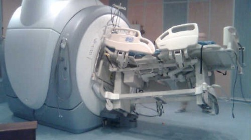
Two years ago, an abstract was presented at this meeting describing the concept of the “hybrid ER.” Check it out using this link. This concept was pioneered in Japan, and consists of a special trauma resuscitation room in the ED with everything but an operating room built into it. It’s possible to perform whole-body CT scan, interventional procedures, and REBOA without moving the patient. Here’s a picture from that abstract:

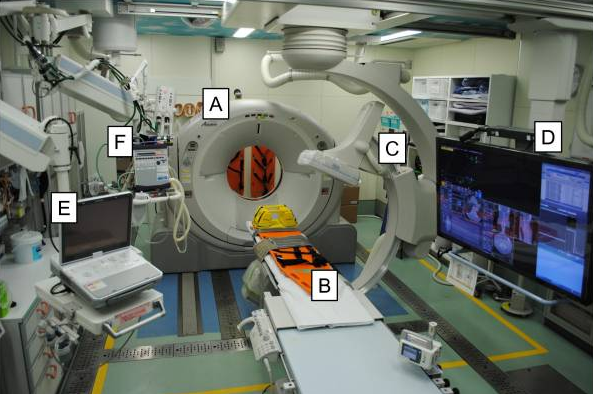
A = CT scanner   B = CT exam table   C = movable C-arm   D = monitor screen   E = ultrasound   F = ventilator

In that abstract, about a thousand patients were compared with two thirds in the hybrid ER group and one third undergoing conventional evaluation. The authors concluded that mortality was significantly improved in the hybrid ER group, and even more so in high ISS patients.

I had a lot of questions for that abstract that were answered in the subsequently published manuscript (reference 2). The authors have updated their experience using new data from the last five years. They created a new approach to resuscitation that is different than the usual ATLS sequence for select patients. Here’s the algorithm they used:

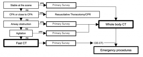
The primary survey is completed, then the patient undergoes a quick whole body CT scan. After that, the secondary survey progresses and any necessary emergency procedures are performed.

In this abstract, the authors compared a group of 46 patients who underwent standard ATLS evaluation with 49 who received the expedited process, which they termed CT First Resuscitation (CTFR). All patients had presumptive hemorrhagic shock based on prehospital vital signs. The authors analyzed injury patterns, interventions performed, timing, adverse events, and outcome. Demographics and injury severity were similar in the two groups.

Here are the factoids:

  • Time to CT in the CTFR group was significantly shorter (1.5 min vs 15 min)
  • The expedited scan settings for CTFR resulted in blindingly fast scan times (median 56 seconds)
  • None of the CTFR patients decompensated during the scan process
  • There was no difference in mortality between CTFR and standard evaluation (14% vs 4%, p=.1)
  • There was no difference in time to hemostatic intervention (56 vs 59 minutes)
  • There was no difference in red cell transfusions (no units in either group)

The authors concluded that CTFR expedited trauma management without adverse effects, and there was no increase in mortality. They, or course, recommended further study.

Bottom line: Several trauma surgeons from a variety of centers wrote an invited commentary last year (reference 3) expressing their excitement about this concept. Reducing time to definitive control of hemorrhage has been repeatedly shown to improve survival. The hybrid ER is one way of reducing those times by eliminating most of the time needed to move the patient about and providing everything but an operating room in the emergency department.

But they also recognized the limitations of this concept. The changes to the ED physical plant are extreme and involve the installation of very expensive equipment that must be heavily shielded from the rest of the emergency department. There are also significant differences in physician training and hospital reimbursement between Japan and the US. This will probably severely limit the adoption of this technology in the States.

I believe that this is an important study showing the feasibility of this method of evaluation. Unfortunately, it does not allow us to draw any real conclusions about safety and efficacy due to the low numbers of patients enrolled. I agree with the authors that a larger study should be performed so we can truly determine whether this concept can possibly be applied outside of Japan.

Here are my questions for the authors / presenter:

  1. Did you perform a power analysis? I doubt that the sample size reported would allow for any findings of statistical significance with the exception of huge differences like time to CT.
  2. How do you protect the trauma team from radiation exposure? Since these patients are in shock when they arrive, I assume that the team cannot leave the room. CT scan radiation exposure of the team is significantly higher than a chest and pelvis x-ray. Repeated team exposure may pose risks.
  3. Does the trend toward higher mortality in the CTFR group trouble you? Sure, it is not statistically significant. But it is approaching significance with a small sample group.
  4. Why didn’t the CTFR group have more rapid hemostatic intervention? One would think these early results could help move the patient to an OR more quickly. And why did it take an hour? Isn’t that a long time?
  5. Why didn’t your patients receive any blood? Weren’t they supposed to be at risk for hemorrhagic shock? How did you treat it without blood? Perhaps your selection criteria need to be tweaked.

This is a nice follow on study from the previous presentation two years ago. It could be an exciting advance in resuscitation, but we need much more info to pass judgement. I’m looking forward to the presentation.

References:

  1. COMPUTED TOMOGRAPHY FIRST RESUSCITATION WITH HYBRID EMERGENCY ROOM FOR SEVERELY INJURED PATIENTS. Plenary paper #25, AAST 2022.
  2. Hybrid emergency room shows maximum effect on trauma resuscitation when used in patients with higher severity. J Trauma Acute Care Surg. 2021 Feb 1;90(2):232-239.
  3. Time to Hemorrhage Control in a Hybrid ER System: Is It Time to Change? Shock. 2021 Dec 1;56(1S):16-21.
Print Friendly, PDF & Email

MRI And External Fixators

MRI is an indispensable tool for evaluation of spine and soft tissue trauma. However, a great deal of effort was be made to ensure that any patient scheduled for this test is “MRI compatible.” The fear is that any retained metallic fragments may move or heat up once the magnets are activated.

But what about trauma patients with external fixators? That is one big hunk of metal that is inserted deep into your patient. There are three major concerns:

  • Is the material ferromagnetic? If so, it will move when the magnets are activated and may cause internal injury. These days, there are many fixator sets that are not ferromagnetic, avoiding this problem.
  • Can currents be induced in the material, causing heating? This is not much of a problem for small, isolated objects. However, external fixators are configured in such a way that loops are created. The fluctuating magnetic fields can induce currents that in turn will heat the surrounding tissue. And thinner materials (narrow pins) result in more current and more heating.
  • Will the metal degrade image quality?

The biggest challenge is that there is no standard ex-fix configuration. Our orthopaedic colleagues get to unleash their creativity trying to devise the appropriate architecture to hold bones together so they can heal properly. This makes it difficult to develop standardized guidelines regarding what can and can’t go into the scanner.

However, there is a growing body of literature showing that the heating effects are relatively small, and get smaller as the distance from the magnet increases. And non-ferromagnetic materials move very little, if at all, and do not interfere with the image. So as long as nonferromagnetic materials are used, the patients are probably safe as long as basic principles are adhered to:

  • Other diagnostic options should be exhausted prior to using MRI.
  • Informed consent must be obtained, explaining that the potential risks are not completely understood.
  • The fixator must be tested with a handheld magnet so that all ferromagnetic components can be identified and removed.
  • All traction bows must be removed.
  • Ice bags are placed at all skin-pin interfaces.
  • The external fixator must remain at least 7cm outside the bore at all times.

Bottom line: MRI of patients with external fixators can be safely accomplished. Consult your radiologists and physicists to develop a policy that is specific to the scanners used at your hospital. 

Related posts:

Print Friendly, PDF & Email

Maxillofacial CT Scans In Children

Facial trauma is common, especially in children. And the use of CT scan is even more common, unfortunately for children. What happens when these two events meet?

I’ve noted that many trauma professionals almost reflexively order a face CT when they see any evidence of facial trauma. This ranges from obvious deformity to lacerations to mere contusions. This seems like overkill to me, since most of the face (excluding the mandible) is visualized with the head CT that nearly always accompanies it.

Finally, someone has actually examined the usefulness of the facial CT scan! The trauma group at Albany collaborated with four other Level I trauma centers, performing a retrospective chart and database review of children (defined as less than 18 years old) who underwent both head and maxillofacial CT scans over a five year period. They excluded penetrating injuries and bites. The concordance of facial fractures seen on head CT vs face CT was evaluated.

Here are the factoids:

  • A total of 322 patients with facial fractures was identified, and the most common mechanisms were MVC, pedestrian struck, and bicycle crash
  • Fractures on head CT matched the facial CT in 89% of cases
  • Of the 35 discordant cases, 21 of the head CTs missed nasal fractures, 9 mandibular fractures, 3 orbital fractures, and 2 maxillary fractures
  • Of those 35 cases, only 7 required operative intervention: 6 mandible fractures and 1 maxillary fracture

The authors concluded that the use of head CT alone with a good clinical exam detects nearly all facial fractures requiring repair.

Bottom line: Although this study confirms my own personal bias and experience, it suffers from the usual problems associated with retrospective studies and small numbers. Nonetheless, the results are compelling. This study provides a way to identify nearly all significant fractures while minimizing radiation to the ocular lens, thyroid, and bone marrow.

The key is a good physical exam, as usual. Inspection of the teeth, occlusion testing, and manipulation of the mandible and maxilla should identify nearly all fractures that might require operation.

Once the exam is complete, a standard head CT should be obtained. Identification of displaced fractures on the head CT should prompt a consult to your friendly facial surgeon to see if they really need additional imaging to determine if the fracture requires operation. Frequently, the head CT images are sufficient and nothing further is required.

Here is the algorithm the authors recommend. Although designed for children, it should work for adults just as well.

Reference: Clinical and radiographic predictors of the need for facial CT in pediatric blunt trauma: a multi-institutional study. Trauma Surg Acute Care Open 2022;7:e000899.

Print Friendly, PDF & Email

The Role Of Postop CT Scan In Penetrating Trauma

CT scans are commonly used to aid the workup of patients with blunt trauma. They are occasionally useful in penetrating trauma, specifically when penetration into a body cavity is uncertain and the patient has no hard signs that would send him or her immediately to the operating room.

Is there any role in operative penetrating trauma, after the patient has already been to the OR? The dogma has always been that the eyeballs of the surgeon in the OR are better than any other imaging modality. Really? The surgical group at San Francisco General addressed this question by retrospectively reviewing 6 years of their operative penetrating injury registry data. They were interested in finding how many occult injuries (seen with CT but not by the surgeon) were found on a postop CT. A total of 225 patients who underwent operative management of penetrating abdomen or chest injury were included. Here are the factoids:

  • Only 110 patients had a postop CT scan; 73 had scans within the first 24 hours, the other 37 were scanned later
  • Rationale for early scan was to investigate retroperitoneal injury in half of patients, but frequently no indication was given (41%)
  • Rationale for late scan was for workup of ileus in one third, or for evaluation of new or unexpected clinical problems
  • Occult injuries were found in about half of early CT patients (52%), and 22% of late CT patients
  • The most common occult injuries were fractures, GU issues, regraded solid organ injury, and unrecognized vascular injuries
  • Ten patients had management changes, including:
    • Interventional radiology for four injuries with extravasation
    • Operation for orthopedic or GU injury in seven patients
    • One patient underwent surgery for an unstable spine fracture

Bottom line: There appears to be a significant benefit to sending some penetrating injury patients to CT in the early postop period. Specifically, those with injury to the retroperitoneum, deep into the liver, near the spine, or with multiple and complicated injuries would benefit. Simple stabs and gunshots that stay away from these areas/structures probably do not need followup imaging. 

Reference: Routine computed tomography after recent operative exploration for penetrating trauma: What injuries do we miss? J Trauma 83(4):575-578, 2017.

Print Friendly, PDF & Email

The Value Of Reinterpreting Outside CT Scans

Okay, one of your referring hospitals has just transferred a patient to you. They diligently filled out the transfer checklist and made sure to either push the images to your PACS system or include a CD containing the imaging that they performed. For good measure, they also included a copy of the radiology report for those images.

Now what do you do?

  • Read the report and consider the results
  • Look at the images yourself and make decisions
  • Have your friendly neighborhood radiologist re-read the images and produce a new report

Correct answer: all of the above. But why? First, you can get a quick idea of what another professional thought about the images, which may help you think about the decisions you need to make.

And one of the few dogmas that I preach is: “read the images yourself!” You have the benefit of knowing the clinical details of your patient, which the outside radiologist did not. This may allow you to see things that they didn’t because they don’t have the same clinical suspicion. Besides, read the images often enough and you will get fairly good at it!

But why trouble your own radiologist to take a look? Isn’t it a waste of their time? Boston Children’s Hospital examined this practice in the context of taking care of pediatric trauma patients. This hospital accepts children from six hospitals in the New England states. In 2010, they made a policy change that mandated all outside images be reinterpreted once the patient arrived. They were interested in determining how often there were new or changed diagnoses, and what the clinical impact was to the patient. They focused their attention only on CT scans of the abdomen and pelvis performed at the referring hospital.

Here are the factoids:

  • 168 patients were identified over a 2-year period. 70 were excluded because there was no report from the outside hospital (!), and 2 did not include the pelvis.
  • Reinterpretation in 28% of studies differed from the original report (!!)
  • Newly identified injuries were noted in 12 patients, and included 7 solid organ injuries, 3 fractures, an adrenal hematoma, and a bowel injury. Three solid organ injuries had been undergraded.
  • Four patients with images interpreted as showing injury were re-read as normal
  • Twenty of the changed interpretations would have changed management

Bottom line: Reinterpretation of images obtained at the outside hospital is essential. Although this study was couched as pediatric research, the average age was 12 with an upper limit of 17. Many were teens with adult physiology and anatomy. There will be logistical hurdles that must be addressed in order to get buy-in from your radiologists, such as how they can get paid. But the critical additional clinical information obtained may change therapy in a significant number of cases.

Reference: The value of official reinterpretation of trauma computed tomography scans from referring hospitals. J Ped Surg 51:486-489, 2016.

Print Friendly, PDF & Email