Category Archives: General

Subdural Hematomas and Hygromas Simplified

There’s a lot of confusion about subdural pathology after head trauma. All subdural collections are located under the dura, on the surface of the brain. In some way they involve or can involve the bridging veins, which are somewhat fragile and get more so with age.

Head trauma causes a subdural hematoma by tearing some of these bridging veins. Notice how thick the dura is and how delicate the bridging veins are in the image below.

When these veins tear, bleeding ensues which layers out over the surface of the brain in that area. If the bleeding does not stop, pressure builds and begins compressing and shifting the brain. A subdural hematoma is considered acute from time of injury until about 3 days later. During this time, it appears more dense than brain tissue.

After about 3-7 days, the clot begins to liquefy and becomes less dense on CT. Many hematomas are reabsorbed, but occasionally there is repeated bleeding from the bridging veins, or the hematoma draws fluid into itself due to the concentration gradient. It can enlarge and begin to cause new symptoms. During this period it is considered subacute.

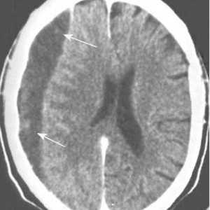
It moves on to a more chronic stage over the ensuing weeks. The blood cells in it break down completely, and the fluid that is left is generally less dense than the brain underneath it. The image below shows a chronic subdural (arrows).

Hygromas are different, in that they are a collection of CSF and not blood. They are caused by a tear in the meninges and allow CSF to accumulate in the subdural space. This can be caused by head trauma as well, and is generally very slow to form. They can lead to slow neurologic deterioration, and are often found on head CT in patients with a history of falls, sometimes in the distant past. CT appearance is similar to a chronic subdural, but the density is the same as CSF, so it should have the same appearance as the fluid in the ventricle on CT.

Related posts:

Print Friendly, PDF & Email

Trauma MedEd Newsletter Released To Subscribers This Weekend!

The April Trauma MedEd Newsletter will be released to subscribers Sunday. This month’s theme is “ED Stuff.” Articles include:

  • Travails of the electronic trauma flow sheet
  • Designing your trauma team
  • Dressing your trauma team
  • Trauma activation levels and triage criteria
  • And more!

Anyone on the subscriber list as of noon (CST) Sunday will receive it that night. Everybody else will have to wait for me to release it here at the end of next week. So sign up for early delivery now by clicking here!

Pick up back issues here!

Print Friendly, PDF & Email

Technology: Airbags For Motorcyclists?

A manufacturer of high-end protective gear for motorcycle and auto racing has developed a fully self-contained airbag system that can be worn. The airbag is easily zippered and connected into a compatible outer jacket, made only by Alpinestars (of course). The airbags expand in much the way that auto airbags do, and they offer protection to the back, shoulders, kidneys, chest, and upper abdomen. A set of colored LEDs on the left sleeve show the rider the status of the system at all times.

A built-in microprocessor samples a set of accelerometers 500 times per second. If any linear or rotational force occurs that is outside of allowed parameters, the airbags inflate well before any impact to the torso occurs. The system is powered by a rechargeable battery that allows for about 25 hours of riding time between charges.

Will this be adopted by the general riding public? Probably not in the near future. The airbag system costs over US $2000, and requires service every two years (US $225). And if it deployed? Another US $550 to repack it. But it’s just a matter of time before similar protective devices worn under a riding jacket make their way to the market at an affordable price.

Related posts:

Print Friendly, PDF & Email

REBOA: All It’s Cracked Up To Be?

Resuscitative endovascular balloon occlusion of the aorta (REBOA) is all the rage. Trauma professionals attending meetings and update courses can count on an update on this interesting technology. But what is it, exactly, and does it work?

REBOA has been around in one form or another for 60 years! In theory, there are five steps for using this technique. 

  1. Access a femoral artery using Seldinger technique
  2. Insert a balloon catheter and move it into position proximal  to the suspected aortic injury
  3. Inflate the balloon to decrease blood loss at the site of injury
  4. Once the injury has been addressed in the OR, slowly deflate the balloon
  5. Then remove it

It’s basically a way of cross-clamping the aorta within the abdomen noninvasively before the patient is opened in the OR. Here’s a diagram that shows what this looks like. Simple, right?

The authors of a recent paper performed an epidemiologic study analyzing data from the Japan Trauma Data Bank over an 8 year period. They performed some sophisticated analyses to try to reduce the usual issues that occur when perusing typical trauma data bank data.

Here are the factoids:

  • Over 45,000 patient records were reviewed, and 452 were included in the study. This is a very large number, as relatively few centers use this technique.
  • The REBOA patients were very badly injured, with a median ISS of 35 and an overall high mortality (76%)
  • The non-REBOA matched patients were less severely injured, with a median ISS of 13 and a 16% mortality
  • When matched for probability of survival using TRISS methodology, the REBOA patients had a significantly higher mortality

Bottom line: What does this mean? Basically, that there is an association with higher mortality given similar injury severity and physiologic compromise, in Japan. The study is another piece in the jigsaw puzzle, and not a good one. Sure, things may be done differently in other countries. And the use of REBOA as a “last ditch effort” certainly may result in higher mortality. But it may not be all it’s cracked up to be. Any use of this technique should be critically evaluated, preferably as part of a well designed study. It’s not for the unprepared or faint of heart.

Reference: Survival of severe blunt trauma patients treated with resuscitative endovascular balloon occlusion of the aorta compared with propensity score-adjusted untreated patients. J Trauma 78(4):721-728, 2015.

Print Friendly, PDF & Email

EAST Guidelines: Blunt Traumatic Aortic Injury

The Eastern Association for the Surgery of Trauma (EAST) has been helping trauma professionals through the publication of practice guidelines for more than 15 years. Members of EAST donate their time to review reams of literature, good and bad, to try to determine the answers to common or puzzling clinical questions.

Why follow a practice guideline? Quite simply, if properly developed, a guideline represents our best understanding of the “correct” answer to the question posed. And as many of you who follow this blog already know, things that “seem to make sense” frequently are totally wrong. Your own experience is poignant, but the pooled experience of the many others who contributed to research on the topic in question is much more significant.

So on the the practice guideline for blunt traumatic aortic injury (BTAI). This one answers three questions. I will list each, followed by the conclusions reached through the literature review.

1. In patients with suspected BTAI, which diagnostic modality should be chosen: CT angiography of the chest, or conventional catheter angiography?

  – Catheter angiography was the standard for decades. When the first EAST guideline on this topic was released 15 years ago, CT angiography was only a level III recommendation because experience with it was lacking. CT technology has advanced rapidly, with multiple detectors, helical scanning, and incredible computing power. Although the quality of the evidence is somewhat low, the panel strongly recommends the use of CT angiography due to its ready availability, speed, low invasiveness, and ability to detect and define other injuries.

2. Should endovascular or open repair be selected in order to minimize stroke, renal failure, paraplegia, and death?

  – Once again, the quality of available data is so so. However, it was possible to detect differences in outcome in comparative studies. The panel strongly recommends the use of endovascular repair in patients who do not have contraindications due to its lower blood loss, mortality, and paraplegia, and equivalent risk of stroke. Furthermore, it is performed more frequently now than open repair, and experience is thus greater at many institutions, further reducing complications.

3. Should the repair be performed immediately or delayed in order to minimize stroke, renal failure, paraplegia, and death?

  – Literature review revealed that the incidence of renal failure and paraplegia were lower with delayed repair, while renal failure was the same in patients with significant associated injuries. There was benefit to delaying repair until resuscitation was achieved and any other life threatening injuries were addressed. The panel recommends that delayed repair be carried out once these other conditions have been corrected. The procedure should not be delayed until the next morning for the convenience of the surgeons.

Related posts:

Reference: Evaluation and management of blunt traumatic aortic injury: a practice guideline from the Eastern Association for the Surgery of Trauma. 78(1):136-146, 2015.

Print Friendly, PDF & Email