Category Archives: General

Heading to SMACC Chicago!

This meeting starts tomorrow at McCormick Place in Chicago! I’ll be speaking tomorrow in Concurrent Session 6: Trauma at 2:55pm. I’ll also be participating in Concurrent Session 16: It’s A Knockdown, a fun panel at 2:30pm on Thursday with the likes of Cliff Reid, Karel Habig, John Hinds, Karim Brohi, Deb Stein, Bill Knight and Andrew Dixon.

Look me up at the meeting or at the opening reception! I look forward to seeing you!

Info on SMACC: click here

View or download the program here

Michael

Print Friendly, PDF & Email

Incidental Pulmonary Embolus

In my last post, I described a case where a fresh trauma patient was found to have an incidental finding of small, distal pulmonary embolus (PE) on her initial trauma evaluation. What should you do when you see this? Reflexively anticoagulate for months?

There are only a few papers dealing with this topic. One is from the MGH, which looked at their experience in screening for deep venous thrombosis (DVT) with duplex ultrasound and diagnosing PE with chest CT. They found that quite a few PEs were found that had no associated DVT in the legs or with clot in the pelvic veins. They also noted an interesting distribution: PEs with no DVT tended to be more distally located, and vice versa for those with DVT. This suggested that some PEs may not be emboli at all, but clot that forms spontaneously in the distal lung circulation.

Scripps Hospital in San Diego did some similar work. Only 31 of some 12,000 patients developed PE, and 19 of these had no identifiable DVT as a source. They also noted that these “de novo” PEs tended to be single and peripherally located. PE associated with DVT tended to be multiple and more central. They also noted an association with chest trauma (pulmonary contusion, rib fractures), blood transfusion, and pneumonia.

Bottom line: As usual, the literature is of little help in this relatively recently identified phenomenon. So what’s the trauma professional to do? Here’s my take. If a PE is found incidentally on the initial trauma evaluation, take a good history to see if there are any family members with clotting problems. Failing that, search for DVT using duplex ultrasound. If the PE is central or multiple, or there is a positive history or duplex screen, anticoagulate as you would any other patient with this problem. If not, carry out the usual prophylaxis and screening as laid out in your usual protocol (you have one, don’t you?), but don’t consider it a “real” PE. At least until we know more about this phenomenon.

Related posts:

Print Friendly, PDF & Email

What Would You Do? Incidental Pulmonary Embolus

You recently received a trauma activation patient after a high speed car crash. She was restrained with belts and airbags, and really has minimal trauma. There is clinical evidence of a few right-sided rib fractures, and nothing else. She has no significant past medical history, but is overweight to obese. You estimate the BMI as about 31.

Following your blunt trauma imaging protocol, one of the scans you obtain is a chest CT. It confirms that the aorta has not been injured and shows two rib fractures. After review, the radiologist calls you with a puzzling result. The patient has a small pulmonary embolus seen in a distal (third order) branch of the pulmonary artery in the left lung.

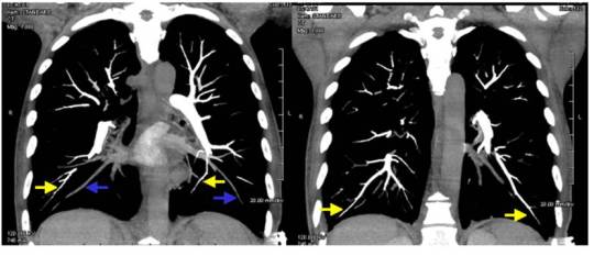
Here’s a coronal view of the scan that you looked at. The arrows show some peripheral branches, but you didn’t see anything on your average resolution monitor.

image

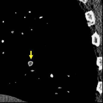
Here’s a sagittal view in high-rez that the radiologist looked at.

image

Yup, looks like a pulmonary embolus. Here are my questions:

  • Where did it come from?
  • What do you do next?
  • What type of treatment is needed?

Think this over this weekend. Discussion and answers on Monday.

Print Friendly, PDF & Email

CT Angiography Of The Head In TBI?

Trauma professionals rely heavily on diagnostic tests these days, particularly CT after blunt trauma. Apparently, the neurosurgeons at the Massachusetts General Hospital are asking for CT angiography of the brain on occasion in patients with TBI. Ostensibly, this is to rule out cases when a brain aneurysm causes a car crash or other blunt trauma.

WTF? Now, I know that we occasionally agonize over older victims of blunt trauma who come in pre-terminal or in arrest. Did they have an MI which caused the event, or did the trauma stop their heart? I had no idea that a ruptured/rupturing aneurysm was such a problem in blunt trauma.

So the surgeons at the MGH decided to critically look at this issue to see if the extra head scan was warranted. 

Here are the factoids:

  • 600 patients with blunt TBI over a one year study period were reviewed
  • 22% underwent CT angio in addition to the normal head CT
  • 66% had the CT angio immediately, 27% within 24 hours, and 7% beyond 24 hours after arrival
  • Specialists who requested the study were neurosurgeons (23), radiologists (15!), neurologists (7!!)
  • Reasons for getting the study: look for cause of subarachnoid hemorrhage (aneurysm) in 43, look for vascular injuries near a skull fracture in 14, rule out stroke in 4, and no particular reason in 71
  • Head CTA changed management in only 1 patient, prompting a formal angiogram which was negative
  • 33 patients (25%) had incidental findings on CTA, but none required any intervention in the hospital or on later followup

Bottom line: There is no value to adding CT angio of the head to the usual scan protocols. Having said that, if the patient was witnessed to lose consciousness prior to the event, and the CT shows subarachnoid hemorrhage in a more typical middle cerebral artery distribution, you might consider it to look for an aneurysm. That’s a lot of if’s. Just move the zebra off the CT scanner first. And as you can see from the last factoid above, if we scan it, we will find stuff. Fortunately, most of that stuff doesn’t need further workup or treatment.

Related posts:

Reference: Is CT angiography of the head useful in management of traumatic brain injury? J Am Col Surg 220(6):1027-1031, 2015.

Print Friendly, PDF & Email