Category Archives: General

Dysphagia and Cervical Spine Injury

Cervical spine injury presents a host of problems, but one of the least appreciated ones is dysphagia. Many clinicians don’t even think of it, but it is a relatively common problem, especially in the elderly. Swallowing difficulties may arise for several reasons:

  • Prevertebral soft tissue swelling may occur with high cervical spine injuries, leading to changes in the architecture of the posterior pharynx
  • Rigid cervical collars, such as the Miami J and Aspen, and halo vests all force the neck into a neutral position. Elderly patients may have a natural kyphosis, and this change in positioning may interfere with swallowing. Try extending your neck by about 30 degrees and see how much more difficult it is to swallow.
  • Patients with cervical fractures more commonly need a tracheostomy for ventilatory support and/or have a head injury, and these are well known culprits in dysphagia

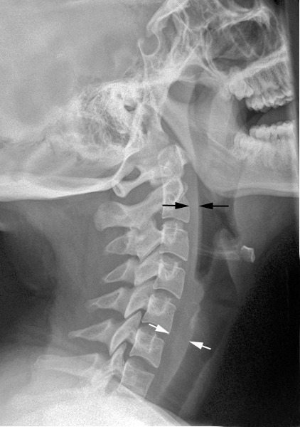
Normal soft tissue (<6mm at C2, <22mm at C6)

A study in the Jan 2011 Journal of Trauma outlined the dysphagia problem seen with placement of a halo vest. They studied a series of 79 of their patients who were treated with a halo. A full 66% had problems with their swallowing evaluation. This problem was associated with a significantly longer ICU stay and a somewhat longer overall hospital stay.

Bottom line: Suspect dysphagia in all patients with cervical fractures, especially the elderly. We don’t use halo vests very often any more, but cervical collars can exacerbate the problem by keeping the neck in an unaccustomed position. Carry out a formal swallowing evaluation, and adjust the collar (or halo) if appropriate.

Reference: Swallowing dysfunction in trauma patients with cervical spine fractures treated with halo-vest fixation. J Trauma 70(1):46-50, 2011.

Print Friendly, PDF & Email

What Percent Pneumothorax Is It?

Frequently, radiologists and trauma professionals are coerced into describing the size of a pneumothorax seen on chest xray in percentage terms. They may something like “the patient has a 30% pneumothorax.”

The truth is that one cannot estimate a 3D volume based on a 2D study like a conventional chest xray. Everyone has seen the patient who has no or a minimal pneumothorax on a supine chest xray, only to discover one of significant size with CT scan.

Very few centers have the software that can determine the percentage of chest volume taken up with air. There are only two percentages that can be determined by viewing a regular chest xray: 0% and 100%. Obviously, 0% means no visible pneumothorax, and 100% means complete collapse. Even 100% doesn’t really look like 100% because the completely collapsed lung takes up some space. See the xray at the top for a 100% pneumothorax.

If you line up 10 trauma professionals and show them a chest xray with a pneumothorax, you will get 10 different estimates of their size. And there aren’t any guidelines as to what size demands chest tube insertion and what size can be watched.

The solution is to be as quantitative as possible. Describe the pneumothorax in terms of the maximum distance the edge of the lung is from the inside of the chest wall, and which intercostal space the pneumothorax extends to. So instead of saying “the patient has a 25% pneumo,” say “the pneumothorax is 1 cm wide and extends from the apex to the fifth intercostal space on an upright film.”

Print Friendly, PDF & Email

EAST 2016: Pain And Tourniquet Efficacy

Ischemia hurts. And tourniquets induce ischemia
on purpose. So logically, tourniquet application should hurt. In a hospital
setting, Doppler ultrasound is used to confirm loss of arterial inflow to the
extremity. In the field, the usual end point is cessation of bleeding. The idea
is to stop tightening the moment that bleeding stops. Unfortunately, this is
not very exact. So the next question is, can pain after tourniquet application
be used to predict how well it is working?

The group at Cook County in Chicago measured
pressures, arterial occlusion, and pain in various extremities in a group of
healthy volunteers (!!). Fortunately for them, complete occlusion was only
maintained for a minute.

Here are the factoids:

  • Three tourniquet systems were used: an
    in-hospital pneumatic tourniquet, the CAT™, and the SWAT™
  • Readings were taken on left and right upper
    arms, the forearms, legs, and the right thigh
  • Using a pain scale of 0-10, tourniquet
    application did not generally induce severe pain
  • Pain scores were 1-3 in the upper arms and forearms,
    3-4 in the thigh, and 2-3 in the leg

Bottom
line: Strangely enough, tourniquet application did not produce severe pain in
any of the subjects. Thigh application tended to be more painful. But,
generally speaking, pain cannot be used as an indicator of effective
application. In the field, cessation of bleeding is the best indicator. And in
the hospital, Doppler ultrasound confirmation should be the standard. In any
case, if the patient is experiencing undue pain after application, check the tourniquet and its positioning.
Something else might be wrong!

Reference:
Pain is an accurate predictor of tourniquet efficacy. EAST 2016 Poster abstract
#23.

Print Friendly, PDF & Email

EAST 2016: Nursing Interruptions In The SICU

A few papers have been published in the nursing
literature about the detrimental effects of interruptions experienced during
patient care. Unfortunately, these papers have never taken the next step to
determine why they occur, and what steps can be taken to decrease the frequency
of this problem.

A group at Wright State in Dayton OH tried to
tease apart the various aspects of this issue. They observed registered nurses
in a 23 bed SICU at a Level I trauma center. A total of 25 sessions covering 75
hours and multiple nurses were analyzed for the cause and duration of any interruption,
and whether it caused a switch from their primary task.

Here are the factoids:

  • Nurses were interrupted every 18
    minutes
    on average
  • The dominant location was in the patient room (58%), and the most
    common activity interrupted was documentation
  • Interruption by an attending or resident was less frequent (10%), but
    ended up being longer than interruptions by other nurses (3 mins vs 1 min)
  • Interruptions of longer duration more commonly
    caused the nurse to switch tasks

Frequency (left) and duration
(right) of interruptions from each source. CL = call light, ECD = electronic
communication device

Bottom
line: This is a first look at the anatomy of nursing interruptions in the SICU.
They are much more common than you think. Task switching (either mentally or
physically) is something that humans do poorly. It always degrades performance,
and can ultimately lead to patient harm. Hopefully, operational protocols can
be developed to protect nurses from unnecessary or non-urgent interruptions to
improve quality of care.

Reference:
The anatomy of nursing interruptions in a surgical intensive care unit at a
trauma center. EAST 2016 Poster abstract #18.

Print Friendly, PDF & Email

EAST 2016: Use Of Scribes To Increase Charge Capture

Ugh! What have we come to? And don’t get me
started regarding the electronic medical record. It is true, the burden of documenting what we do in
order to get paid continues to increase
. And, of course, this takes time.
And we don’t get paid for the time we spend doing this documentation.

The next
logical step? Hire cheap labor to do the documentation!
This is becoming a
fairly common practice, unfortunately. The next abstract is from MetroHealth in
Cleveland. They looked at the impact of hiring scribes from both time and money
standpoints. Two five-month periods were reviewed, with and then without
scribes. Hold on, the numbers here will make your head spin!

Here are the factoids:

  • A total of 9726 notes were written in the no-scribe period, and 10933
    were written with scribes
  • Despite the fact that there were 407 fewer patient-days during the
    scribe period, 882 more inpatient notes were written
  • More progress notes were written early in the day with scribes, and
    fewer were written during the evening hours
  • The number of notes written after patient discharge decreased from
    12.7% to 8.4% when using scribes
  • Charge capture increased by $32 per patient-day (?!)
  • The additional scribe notes resulted in an extra
    $316K in charges generated 

Bottom
line: It’s just so annoying to think that we actually have to stoop to
something like this. The scribes cost this program about $33K. They generated
$316K. The paper estimates that they were actually paid about 20% of this, or
$63,000. So each of the eight trauma surgeons in this program collected an
extra $8,000 over the 5-month period.

So, is it
worth it? Maybe. The extra charges collected seem insignificant. But, if the
surgeon is actually able to dedicate less time to documenting, and this allows
them to spend more time operating (which is what really pays the bills), then
perhaps it is. I’m not easily convinced, though.

Reference:
Trauma surgeons save lives – scribes save trauma surgeons! EAST 2016 Poster
abstract #16.

Print Friendly, PDF & Email