Category Archives: General

Pulmonary Embolism and DVT in Trauma

We have long assumed that pulmonary emboli start as clots in the deep veins of the legs (or pelvis), then break off and float into the branches of the pulmonary artery in the lungs. A huge industry has developed around how best to deal with or prevent this problem, including mechanical devices (sequential compression devices), chemical prophylaxis (heparin products), and physical devices (IVC filters).

The really interesting thing is that less than half of patients who are diagnosed with a pulmonary embolism have identifiable clots in their leg veins. In one study, 26 of 200 patients developed DVT and 4 had a PE. However, none of the DVT patients developed an embolism, and none of the embolism patients had a DVT! How can this kind of disparity be explained?

Researchers at the Massachusetts General Hospital retrospectively looked at the correlation between DVT and PE in trauma patients over a 3 year period. DVT was screened for on a weekly basis by duplex venous ultrsonagraphy. PE was diagnoses exclusively using CT scan of the chest, but also included the pelvic and leg veins to look for a source. A total of 247 patients underwent the CT study for PE and were included in the study.

Here are the factoids:

  • Forty six patients had PE (39% central, 61% peripheral pulmonary arterial branches) and 18 had DVT (16 seen on the PE CT and 2 found by duplex)
  • Of the 46 patients with PE, only 15% had DVT
  • All patient groups were similar with respect to injuries, injury severity, sex, anticoagulation and lengths of stay
  • Interestingly, 71% of PE patients with DVT had a central PE, but only 33% of patients without DVT had a central PE.

The authors propose 4 possible explanations for their findings:

  1. The diagnostics tools for detecting DVT are not very good. FALSE: CT evaluation is probably the “gold standard”, since venography has long since been abandoned
  2. Many clots originate in the upper extremities. FALSE: most centers do not detect many DVTs in the arms
  3. Leg clots do not break off to throw a PE, they dislodge cleanly and completely. FALSE: cadaver studies have not shown this to be true
  4. Some clots may form on their own in the pulmonary artery due to endothelial inflammation or other unknown mechanisms. POSSIBLE

An invited critique scrutinizes the study’s use of diagnostics and the lack of hard evidence of clot formation in the lungs.

Bottom line: this is a very intriguing study that questions our assumptions about deep venous thrombosis and pulmonary embolism. More work will be done on this question, and I think the result will be a radical change in our use of anticoagulation and IVC filters over the next 3-5 years.

Reference: Pulmonary embolism and deep venous thrombosis in trauma: are they related? Arch Surg 144(10):928-932, 2009.

Print Friendly, PDF & Email

More On CT Usage In Unstable Patients

Yes, it is practically dogma that CT should not be used in unstable trauma patients. Either they go directly to the OR, or an attempt to stabilize them is briefly undertaken in the trauma bay. And as you know, I’m not a big believer in dogma. But this one has withstood the test of time. You can see my comments about a previous paper below in the related posts.

But now some authors in Colombia have published a paper that seems to call this idea into question. Could it be true? Read carefully!

This was a small, retrospective review of patients from a large Level I government designated trauma center. They reviewed their experience over a two year period, identifying all hemodynamically unstable patients in the registry. They excluded dead patients, those with isolated head injury, and any who had surgery at an outside hospital prior to transfer.

Here are the factoids:

  • 171 patients were reviewed, and of course they tended to be young males
  • 91 went straight to the OR, and 80 were taken to CT first
  • “Unstable” patients were defined as having SBP < 100 and/or HR > 100
  • Mechanism of injury for the OR group was 95% penetrating, but for the CT group was about 50:50 penetrating/blunt
  • The mean SBP and HR for the “unstable” patients taken to CT were 92 and 110, respectively
  • Mortality was the same for both groups (18% OR vs `13% CT)

Bottom line: The authors concluded that it is permissible to take unstable patients to CT if you don’t spend too much time there based on similar mortality rates. But the problem was that I don’t consider their patients to have been unstable! Mean SBP in their “unstable” group was over 90 torr and the heart rate was only 110! The lowest SBP was only 79. And mortality is too crude of an outcome to rely on. Furthermore, the patients they took to CT tended to have blunt mechanisms, and may not have had ample efforts at resuscitation in the trauma bay first, or may have met criteria to go to CT anyway (see related posts below).

Reference: Computed tomography in hemodynamically unstable severely injured blunt and penetrating trauma patients. J Trauma 80(4):597-603, 2016.

Print Friendly, PDF & Email

What The Heck? Head CT – The Answer

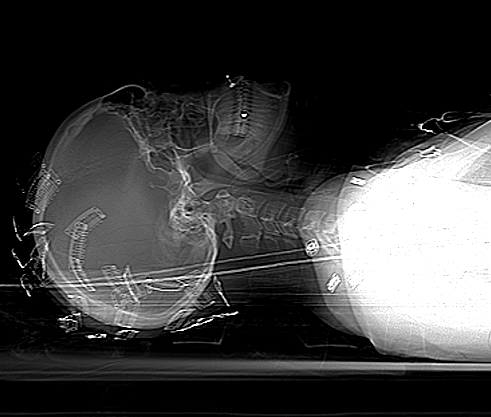
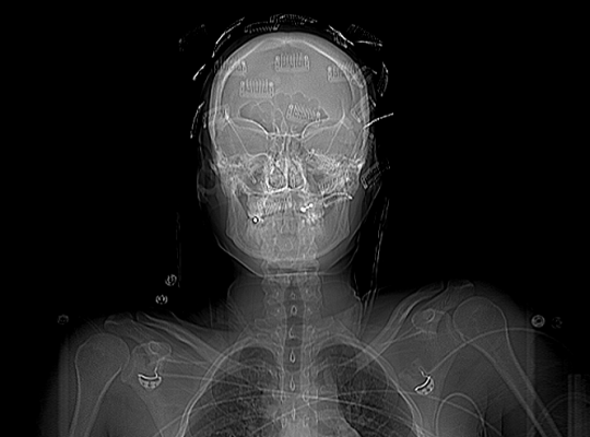
It appears that no one was able to figure this one out! To recap, a young person (female in this case) sustained blunt trauma to the head. When her head was scanned, the following was found on the scout scan:

What is all this odd stuff? They look like some kind of metallic clips placed all over the head. The answer? Hair extensions! Here’s what they look like up close.

Unfortunately, they cannot be left in place during the CT. The amount of scatter is significant enough to degrade the quality of the study. By definition, if you have taken your patient to CT, they are stable and you have a little time. So carefully remove all of the extensions and place them in a bag and save them for the patient (they can be expensive!). 

Make sure the CT tech obtains another topogram to confirm removal of all of the extensions. Then proceed with your CT as usual!

Print Friendly, PDF & Email

Why Do They Call It: Extra-axial Blood?

You’ve seen it on head CT reports. “The patient has a collection of extra-axial blood…” Then it goes on to describe the location and size of a subdural hematoma. But why is it called “extra-axial?”

The answer lies in the embryology of the central nervous system. Yes, it’s been a long time since any of us have read anything about that. Early animals had a straight neural tube, which slowly evolved into a brain and spinal cord. This is known as the axis of the nervous system.

The brains of early vertebrates developed at the end of the neural tube, and were oriented in the same longitudinal axis as the rest of it. As brains got bigger, a 90 degree bend occurred at the cephalic flexure.

So in humans, there is a difference between the body axis and the brain axis. But the brain axis is what really counts. This means that any blood outside of the brain axis is defined as extra-axial.

Bottom line: Extra-axial blood is defined as any bleeding outside of the brain parenchyma. This includes subdural and epidural hematomas, and subarachnoid hemorrhage. It does not include any intraparenchymal bleeding like contusions, strokes, or hematomas.

Print Friendly, PDF & Email