Category Archives: General

How To Troubleshoot Air Leaks in Chest Tube Systems

An air leak is a sure-fire reason to keep a chest tube in place. Fortunately, many air leaks are not from the patient’s chest, but from a plumbing problem. Here’s how to locate the leak.

To quickly localize the problem, take a sizable clamp (no mosquito clamps, please) and place it on the chest tube between the patient’s chest and the plastic connector that leads to the collection system. Watch the water seal chamber of the system as you do this. If the leak stops, it is coming from the patient or leaking in from the chest wall.

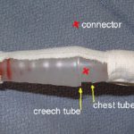
If the leak persists, clamp the soft Creech tubing between the plastic connector and the collection system itself. If the leak stops now, the connector is loose.

If it is still leaking, then the collection system is bad or has been knocked over.

Here are the remedies for each problem area:

  • Patient – Take the dressing down and look at the skin entry site. Does it gape, or is their obvious air hissing and entering the chest? If so, plug it with petrolatum gauze. If not, the air is actually coming out of your patient and you must wait it out.
  • Connector – Secure it with Ty-Rap fasteners or tape (see picture). This is a common problem area.
  • Collection system – The one-way valve system is not functioning, or the system has been knocked over. Click here for an example. Replace it immediately.

Note: If you are using a “dry seal” system (click here for more on this) you will not be able to tell if you have a leak until you fill the seal chamber with some water.

Print Friendly, PDF & Email

DPL: A Dying Art?

Diagnostic peritoneal lavage (DPL) was invented by David Root at this hospital (Ancker Hospital, which then became St. Paul Ramsey, now Regions Hospital) in the 1960’s. It enjoyed its heyday during the 70’s and 80’s, when it was done hundreds of times per year at most major trauma hospitals. Now, we do it about 5 times per year. What happened?

As you know, DPL is a qualitative test. It gives a yes/no answer to the question “does this patient need an operation?” based on red and white blood cell counts. During the mid-1980s, CT scanning was introduced, which provides much more quantitative information about injuries in the abdominal cavity. The improved ability to diagnose abdominal injury, especially solid organ injury, has led to the demise of DPL.

Most solid organ injury results in some free blood in the peritoneal cavity. It doesn’t take much blood (10 cc of whole blood mixed with 1 liter of infused crystalloid) to exceed the threshold of 100,000 RBC per ml of aspirate that will send the surgeon off to the OR. Therefore, pretty much any liver or spleen laceration would have to be taken to the OR based on a DPL.

But we know that very few liver/spleen injuries actually need an operation. So DPL cannot be used, or the negative laparotomy rate for blunt trauma would escalate. The other downside to DPL is that it’s not possible to get all of the infused crystalloid back out of the abdomen. This leads to a confusing amount of free fluid seen on any CT scan done after a DPL.

So DPL is now down but not out. Some practical pointers:

  • DPL should be used primarily as a backup to an equivocal or unbelievable FAST exam in an unstable patient. An example would be a patient who is hypotensive, has a negative FAST and no other obvious bleeding sources.
  • Remember to insert a gastric tube and urinary catheter so the stomach and bladder are decompressed before the procedure. The easiest way to remember this is to tape these catheters to the DPL procedure tray.
  • A DPL is actually 2 procedures: peritoneal tap and lavage. Once the catheter is in, it should be aspirated. If 10cc of gross blood is returned, the test is positive and the patient needs to go immediately to OR.
  • For blunt trauma, the threshold for RBC per µl is 100,000. The threshold for WBC is 500 per µl. If particulate material or weird colors are seen (stool or bile), the test is also considered positive. Send the sample for cell counts only. Don’t send for any other assays (e.g. amylase). 
  • For penetrating trauma, the thresholds have never been well defined. A number around 25,000 RBC per µl probably provides the best balance between sensitivity and negative laparotomy rate.

Reference: Diagnostic peritoneal lavage. HD Root, CW Hauser, CR McKinley, JW LaFave, RP Mendiola Jr. Surgery 57(5):633-637, 1965.

Print Friendly, PDF & Email

When to Give Spleen Vaccines After Splenectomy for Trauma

I’ve written previously on the (f)utility of giving vaccines after splenectomy for trauma (click here to read). However, it is more or less a medicolegal standard, so pretty much everyone gives them. The big question is, when? 

Some centers give them immediately postop, some before hospital discharge, and some during their postop visit. Who is right? The argument is that major surgery produces some degree of immunocompromise. So if the vaccines are given too early, perhaps the anitbodies will not be processed as effectively, and the response to an actual bacterial challenge might not be as good.

One prospective study randomized patients to receive their pneumococcal vaccine either 1, 7, or 14 days after surgery. IgG levels were measured before vaccination and again after 4 weeks. This study found that antibody concentrations were the same in all groups. However, functional activity of the antibodies was low in the 1 and 7 day groups, and nearly normal in the 14 day group.

Following this, a rat study looked at vaccination timing followed by exposure to pneumococcus. These animals were splenectomized, then given a real or sham vaccination at 1, 7, or 42 days. They then had pneumococcus injected into their peritoneal cavity. About 70% of all rats with sham vaccination died. Only 1.5% of the vaccinated rats died, and there were no differences based on vaccination timing.

Bottom line: Neither antibody titer studies nor rat studies easily translate into recommendations for treating overwhelming post-splenectomy sepsis (OPSS) in humans. And such a study can never be done because of the rarity of this condition (less than 70 cases since the beginning of time). It really boils down to your specific population, balancing your assurance that your patient will get it against the possibility that their immune system may not react to it as much as it could. 

At our center, we give the vaccines as soon as possible postoperatively. This ensures that it is given, and erases any doubt of what might happen if the patient does not show up for their postop check.

References:

  • Immune responses of splenectomized trauma patietns to the 23-valent pneumococcal polysaccharide vaccine at 1 versus 7 versus 14 days after splenectomy. J Trauma 44(5):760-766, 1998.
  • Timing of vaccination does not affect antibody response or survival after pneumococcal challenge in splenectomized rats. J Trauma 45(4):682-697, 1998.

Related posts:

    Print Friendly, PDF & Email

    Pneumothorax in Children

    Last week I wrote about pneumomediastinum in children (click here to read). This week I’ll talk about a somewhat more common problem: pneumothorax. This condition is far more mysterious than in adults.

    Sports related pneumothorax rarely occurs without rib fractures, which are themselves uncommon in children. The usual mechanism is barotrauma, most likely from an impact while the glottis is closed. The typical presentation is that of pleuritic chest pain, which may be followed by dyspnea. Focal chest wall tenderness is typically absent. Teenagers tend to engage in more strenuous activity and are more likely to actually sustain a rib fracture, so they may have focal tenderness over the fracture site.

    Spontaneous pneumothorax in children is also uncommon. However, it is a very different entity. It may be related to blebs in the lungs, and may be more common in children who were born prematurely. The recurrence rate after successful treatment is approximately 50% (in small series). Recurrence is not predictable by looking for blebs on chest CT. The recurrence rate is significantly lower after VATS.

    Bottom line: A child who complains of pleuritic chest pain, and especially dyspnea, should undergo a simple PA chest xray. If a pneumothorax is present, consider the following:

    • Insert a small chest tube or catheter if needed, the smaller the better. (I’m stilling looking for the answer to the question of how big a pneumothorax is big enough)
    • Don’t use high inspired oxygen; it doesn’t work. (Read my older blogs from 2010 – this one and this one, too)
    • Don’t get a chest CT for either the initial pneumothorax or any recurrences (too much radiation, too little utility)
    • If this is a spontaneous pneumothorax, caution the parents on the possibility of recurrence
    • If a spontaneous pneumothorax does recur, consult a pediatric surgeon to consider VATS pleurodesis
    • When can the child return to sports? There is absolutely no good literature. I recommend the usual time it takes most soft tissues to return to full tensile strength after injury (6 weeks).

    References:

    • Management of primary spontaneous pneumothorax in children. Clin Pediatr, online ahead of print, April 11, 2011.
    • Sports-related pneumothorax in children. Pediatric Emergency Care 21(4): 259-260, 2005.

    Related posts:

    Thanks to Chris Chow MD for finding some of the literature for this post.

    Print Friendly, PDF & Email

    New Technology II: Helping Paraplegics and Quadriplegics to Walk

    The second company that makes a device to assist walking in spinal cord injured patients is Berkeley Bionics. Their exoskeleton is lighter (45 pounds) and more form-fitting, making it easier to maneuver indoors. It can operate for up to 6 hours between charges. The unit does require operator assistance in the form of a pair of canes for balance.

    Prices were not available for the products from both Berkeley and Rex. However, the technology looks promising for several reasons. It allows the subject to stand upright, putting weight on their feet. This helps increase muscle tone and maintain joint flexibility. It also decreases pressure problems caused by remaining seated.

    These devices are in an early stage right now. As the technology advances, expect to see smaller bionics with better (smoother) computer control, and more access for people with higher spinal cord injuries.

    Disclaimer: I have no financial interest in Berkeley Bionics.

    Print Friendly, PDF & Email