Category Archives: General

Portable CT Scanning For Trauma Patients

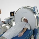
I recently had the opportunity to see a portable head CT scanner in action, the CeroTom by NeuroLogica (Danvers, MA). Today, I’ll give my thoughts on this new technology.

There are 3 major considerations when evaluating portable CT scanning:

  • Patient safety, always at the forefront
  • Usefulness, also know as image quality
  • Financial viability

From a safety standpoint, portable scanning can decrease (but not eliminate) the safety hazards associated with transporting a critically ill patient out of the ICU. Road trips are associated with misplaced/displaced lines, tubes and monitors about 15% of the time. These are lifelines in some patients, and even momentary disruptions can be life-threatening. Some patients are on levels of support so high they are not transportable, so portable scanners offer an opportunity to get diagnostic imaging that would not be available otherwise.

Clinical performance is on par with standard scanners. Resolution is lower, but the diagnostic accuracy and reliability are not different compared to fixed scanners.

From a financial standpoint, use of the portable scanner works as well. The Cleveland Clinic deployed a CereTom scanner a few years ago and found that the unit paid for itself in 6.9 months. For you financial types, the internal rate of return was 169% and the 5-year expected economic benefit was $2.6 million.

Bottom line: This new piece of technology offers significant benefits to patients in the ICU who may otherwise not be able to get imaging due to safety reasons. It can also be employed in the OR on anesthetized patients, which can assist with diagnosis in patients with both abdominal injuries requiring immediate operation and concomitant head injury.

Practical notes: The CereTom is an 8-slice scanner with a 25cm field of view. The patient is moved onto a scan board which supports the head while it is moved slightly off the top of the bed to accommodate the scanner. Current scanner cost is $450,000 and attachment packages for hospital beds are $7,000. One CT technologist can operate the unit, which takes about 5 minutes to set up and 15 minutes to scan. All lines, tubes and monitors must be (carefully) moved to the side of the bed so the scanner can fit over the top.  

References:

  • The economic and clinical benefits of portable head/neck CT imaging in the intensive care unit. Radiology Manage 30(2):50-54, 2008.
  • Review of portable CT with assessment of a dedicated head CT scanner. Am J Neuroradiol 30:1630-1636, 2009.

I have no financial interest in Neurologica, Inc.

Print Friendly, PDF & Email

Rib Fracture Management

A reader sent a query yesterday regarding treatment of rib fractures, and specifically asking about epidural analgesia. Today, I’ll try to answer those questions.

Rib fractures, with or without other injuries, are a big killer in trauma patients. This is particularly true in the elderly. Overall mortality rates range from 3% to 13%, with the most import factor being pain. So what is the best way to manage patients with rib fractures to speed their safe recovery?

It’s best to attack this problem from three different directions simultaneously: pain control, respiratory hygiene (or pulmonary toilet if you’re a pessimist), and activity management.

There are many approaches to pain management, which include:

  • Oral or IV analgesics
  • Various types of blocks (intrapleural, intercostal, paravertebral, epidural)
  • Topical agents (xylocaine patch)
  • Stabilization (surgical only; belts and straps are bad for breathing)

Epidural analgesia is usually seen as the ultimate form of pain control, and is usually recommended for patients with multiple fractures or severe pain with inadequate response to medications and blocks. Much of the literature on its use is based on ICU patients who were not injured. A meta-analysis was conducted that specifically looked at epidural analgesia results in trauma patients, and found that it did improve pain management and some pulmonary function tests. However, there did not appear to be any change in mortality, ICU or hospital length of stay, or time on a ventilator.

Respiratory hygiene may involve simple measures such as coughing and deep breathing, incentive spirometry, and even mechanical ventilation in severe cases. Activity management consists of turning, sitting in a chair, walking, and forms of mechanical chest wall oscillation.

Bottom Line: The key to rib fracture management is a systematic approach that address all three dimensions of care based on objective patient measures. One size does not fit all, so more aggressive measures are warranted for more severe injury. I’ve attached an interesting patented scoring system and management algorithm, as well as two protocols from US trauma centers that range from simple (Vanderbilt) to more complex (West Virginia University).

Please feel free to comment, and I’d be happy to look at your protocol. Please email it to me!

Related post: History of epidural analgesia

Downloads

References

  • Effect of epidural analgesia in patients with traumatic rib fractures: a systematic review and meta-analysis of randomized controlled trials. Can J Anaesth 56(3):230-42, Epub 2009 Feb 11.
  • Rib Fracture Score and Protocol, US Patent #7,225,813 B2 – June 5, 2007
Print Friendly, PDF & Email

Trauma 50 Years Ago! Gunshot Wound Debridement

I’ve generally written a post every month reviewing an article from the Journal of Trauma exactly 20 years earlier that illustrates the history of some of the things we do now. I’m reaching further back in the past today, looking 50 years ago to the July 1961 issue of the first volume of the Journal.

Most trauma hospitals do not see many gunshots. There are exceptions, of course, in more urban areas. Much of what we’ve learned about taking care of gunshot wounds is based on experiences gained from the military during wartime. In the 15 years after World War II, many hospitals were treating civilian gunshot wounds like their military counterparts. 

A paper published in 1961 reported the current practice at a number of trauma hospitals across the US. Remember, there were no “trauma centers” at the time. These reports were from Bellevue in New York, St. Louis, Cook County in Chicago, Galveston, Columbus and others.

A total of 368 wounds were managed, and more than 300 were cared for without the wound debridement that had been the norm. The authors found that most did very well with cleansing and antibiotic treatment. They concluded that debridement was not necessary unless a vascular injury was also present. It was believed that the firearms found in civilian practice were universally low velocity weapons which did not inflict the degree of tissue damage of military weapons.

We generally follow this tenet to this day. Most handgun wounds do not need any special debridement. Rifle, shotgun and assault weapon injuries typically do, and is best carried out in an OR. Antibiotic use has decreased significantly, in many cases to a single dose of a drug that covers typical skin bacteria.

Reference: The indications for debridement of gun shot (bullet) wounds of the extremities in civilian practice. J Trauma 1(4):368-372, 1961.

Print Friendly, PDF & Email

PAs and NPs In Level I Trauma Centers

Trauma service staffing is important to maintaining trauma center status. Teaching centers in the US have been grappling with resident work hour rules, and non-teaching centers have always had to deal with how to adequately staff their trauma service. What is the impact of staffing a trauma center with midlevel practitioners (MLPs) such as physician assistants and nurse practitioners?

A state designated Level I trauma center in Pennsylvania retrospectively examined the effect of adding MLPs to an existing complement of residents on their trauma service. They examined the usual outcomes, including complications, lengths of stay, ED dwell times and mortality. 

Here are the more interesting results:

  • ED dwell time decreased for trauma activations and transfers in, but it increased for trauma consults. Of note, data on dwell times suffered from inconsistent charting.
  • ICU length of stay decreased significantly
  • Hospital length of stay decreased somewhat but did not achieve significance
  • The incidence of most complications stayed the same, but urinary tract infection decreased significantly
  • There was no change in mortality

Bottom line: There is a growing body of literature showing the benefits of employing midlevel providers in trauma programs. Whereas residents may have a variable interest in the trauma service based on their career goals, MLPs are professionally dedicated to this task. This study demonstrates a creative and safe solution for managing daily clinical activity on a busy trauma service.

Reference: Utilization of PAs and NPs at a level I trauma center: effects on outcomes. J Amer Acad Physician Assts, July 2011.

Print Friendly, PDF & Email

Pulmonary Contusion After Sports Injury

Most pulmonary contusions occur after diffuse, high energy blunt trauma like a fall or car crash. Occasionally, pulmonary injury can occur after a much more focal injury like an impact during sports. This may occur due to direct impact from another player, or from a rapidly moving object like a ball or puck. Today, I’ll focus on the latter, impact from a fast-moving object.

Typically, the athlete will complain of pain at the area of impact and some degree of breathing impairment. This is usually due to musculoskeletal pain, rib fracture or involuntary splinting. It is possible to develop pneumothorax or hemothorax, especially if a rib is fractured. At some point, hemoptysis may be present. This is pathognomonic for the presence of a pulmonary contusion.

Any athlete with more than mild to moderate pain, or any physical exam findings other than tenderness, should be more fully evaluated. Here are some important tips:

  • The only imaging required is a two view conventional chest xray. This will identify significant pneumothorax, hemothorax or contusions that require additional management.
  • Chest CT is not indicated. It does not change management.
  • Use your typical algorithms for managing hemothorax and pneumothorax
  • A pneumatocele visible on the xray indicates a small pulmonary laceration. A followup xray or two may be needed to ensure that it does not expand or cause a pneumothorax. Thoracic surgery involvement is not usually indicated
  • Hemoptysis is normal, and may present immediately or several days later. The patient should be reassured in advance so they don’t call you frantically in the middle of the night.

Image source: internet

    Print Friendly, PDF & Email