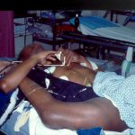
Explain This!
Any idea how this can happen? And what exactly is wrong?
Source: personal archive. Not treated at Regions Hospital

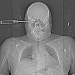
Explain This!
Any idea how this can happen? And what exactly is wrong?
Source: personal archive. Not treated at Regions Hospital
I’ve written several times on the importance of getting patients off the backboard promptly in the ED. Many hospitals use slide boards to facilitate patient movement on and off the ED cart when undergoing imaging studies. How should we manage the use of this device?
There is no difference between a backboard and a slide board to the patient. It’s hard and uncomfortable to lie on for any period of time, and can cause soft tissue injury. To trauma professionals in the ED it is thinner, less bulky, easier to manipulate, and does not interfere with xrays as much. We tend to pay less attention to it than a backboard. Although it does not immobilize the spine as well as a backboard does, the difference is not clinically significant (in a cooperative patient). Remember, if your patient actually has a spine fracture, they will be placed on logroll precautions on a soft mattress only somewhere in your hospital! No stiff boards of any kind!
Slide board management tips:
Related posts:
When Can You Close That Stab Wound?
I find that many trauma professionals are nervous about closing stab wounds. They seem to worry a lot about infections and lean toward leaving the wound open to heal by secondary intention. But is this warranted?
The answer is: probably not. Most knives used for assaults are clean, but not quite sterile. Yes, there are a few bacteria on the blade, but not very many. So if the usual wound management guidelines are followed, the patients generally do quite well.
The guidelines are:
If any of these guidelines have been violated, it’s probably best to leave the wound open. Otherwise the default should be to try to close it as soon and as cleanly as possible. This means irrigating with saline to decrease any bacterial counts. Either sutures or staples are acceptable.
The most important part of this process is patient education. They must be informed about what signs of a wound infection to look for so they can return earlier rather than later to have you deal with it.
Related posts:
Diaphragm injuries are notoriously hard to detect, and there is a significant rate of delayed or missed diagnosis. Today I’ll offer a few practical tips on finding and managing this rare injury.
Mechanism is important. Penetrating injury is more common, and it can be really tough to diagnose this injury in stabs to the lower chest. Anything below the nipples is suspect. Blunt injury requires substantial force. This is seen in deceleration injuries, usually with a crush component to the abdomen. Ejection and partial ejection is common.
Left sided injury is much more common than right. The liver probably diffuses the force more evenly, protecting the right diaphragm. Be very skeptical if a radiologist tries to tell you the patient has a right diaphragm injury.
Patients have significant symptoms with blunt injury. Respiratory distress is common on the left, and deep visceral pain on the right if the liver partially herniates into the chest. Serious associated injuries are common due to the high energy involved.
Diagnosis is difficult. CT is a very good, but imperfect, tool. Coronal and sagittal reconstructions can be very helpful.
If you are inclined to explore for a possible diaphragm injury in a stable patient, consider laparoscopy first. But warn the anesthesiologist! If your patient has the injury, they will rapidly develop a complete pneumothorax, so you need to be ready to quickly insert a chest tube.
Caress the diaphragm to find small holes. Place your hand in the palm up position so your fingers will drop into any defect. Be thorough, since small knife and bullet wounds are easy to miss, especially when located posteriorly.
Related post:
Ten months ago I wrote about getting patients off backboards as soon as possible. The question has arisen again, so I did a little digging to find some good science behind this. And I found it.
This problem has been looked at three ways. From best to worst they are: studies on OR patients who developed pressure ulcers postop, studies on animals, and studies on tissues. I’ll focus on the first because a real person who is chemically and physically restrained to an OR table is very similar to one who has been fastened to a backboard.
The most cited study (retrospective, of course) showed that patients who had tissue pressure over bony prominences that exceeded their diastolic pressure developed pressure ulcers within 6 hours, and even faster with higher tissue pressures. But even better prospective OR studies have been done, and these showed that ulcers could occur in as little as three hours.
Keep in mind that these studies involved patients in whom real efforts were made to pad bony prominences and actively avoid tissue injuries. Yet they still occurred. Contrast this with a patient who is strapped to a hard backboard in your ED, with little ability to adjust their position to improve circulation.
Related work has shown that:
Bottom line: Get your patients off that backboard ASAP! I recommend sliding it out when they are logrolled to examine the back. The board is of little or no benefit to spine stability in a cooperative patient. And we have ways of encouraging cooperation if they are not.
Related post:
Reference: How Much Time Does it Take to Get a Pressure Ulcer? Integrated Evidence from Human, Animal, and In Vitro Studies. Ostomy Wound Management. 54(10):26-8, 30-5, 2008.