Category Archives: General

What The Heck? Part 2: Progressive Back Pain After Heavy Lifting

Yesterday, I described a case of a young athlete who developed progressive back pain after rapidly increased his deadlift weights. He presented to the hospital with back pain and inability to get up from a supine position. He had firm and tender paraspinal muscles in his lower back, but no other findings.

What to do next? Obviously, we need a bit more information on the bony structures. Other than run of the mill muscle strain, a compression fracture would be the next most common diagnosis. In this young, healthy athlete, a simple set of AP and lateral spine images should be sufficient. But if you opted for a CT scan, I won’t argue. In either case, the images were normal.

Since there is significant muscle pain and tenderness, a lab panel with a few extras is in order, as well. The usual electrolytes, etc were normal. Creatinine was 0.9, but CPK was 60,000!

Now what are you thinking? What’s the diagnosis, and what is the decision tree for treatment?

Add your comments below, or tweet them out. I’ll finish this topic up in the next post.


Print Friendly, PDF & Email

Autotransfusing Blood Lost Through The Chest Tube

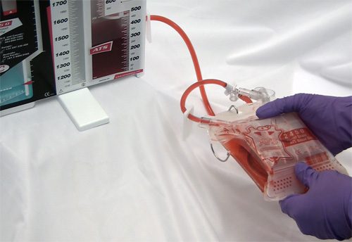
Autotransfusing blood that has been shed from the chest tube is an easy way to resuscitate trauma patients with significant hemorrhage from the chest. Plus, it’s usually not contaminated from bowel injury and it doesn’t need any fancy equipment to prepare it for infusion.

It looks like fresh whole blood in the collection system. But is it? A prospective study of 22 patients was carried out to answer this question. A blood sample from the collection system of trauma patients with more than 50 cc of blood loss in 4 hours was analyzed for hematology, electrolyte and coagulation profiles.

The authors found that:

  • The hemoglobin and hematocrit from the chest tube were lower than venous blood (Hgb by about 2 grams, Hct by 7.5%)
  • Platelet count was very low in chest tube blood
  • Potassium was higher (4.9 mmol/L), but not dangerously so
  • INR, PTT, TT, Factor V and fibrinogen were unmeasurable

Bottom line: Although shed blood from the chest looks like whole blood, it’s missing key coagulation factors and will not clot. Reinfusing it will boost oxygen carrying capacity, but it won’t help with clotting. You may use it as part of your massive transfusion protocol, but don’t forget to give plasma and platelets according to protocol. This also explains why you don’t need to add an anticoagulant to the autotransfusion unit prior to collecting or giving the shed blood!

Reference: Autotransfusion of hemothorax blood in trauma patients: is it the same as fresh whole blood? Am J Surg 202(6):817-822, 2011.

Print Friendly, PDF & Email

Trauma In Pregnancy 5: C-Section – When?

The perimortem C-section (PMCS) is a heroic procedure designed to salvage a viable fetus from a moribund mother. Interestingly, in some mothers, delivery of the fetus results in return of spontaneous circulation.

The traditional teaching is that PMCS should be started within 4-5 minutes of the mother’s circulatory arrest. The longer it is delayed, the (much) lower the likelihood that the fetus will survive.

The reality is that it takes several minutes to prepare for this procedure because it is done so infrequently in most trauma centers. Recent literature suggests the following management for pregnant patients in blunt traumatic arrest (BTA):

  • Cover the usual BTA bases, including securing the airway, obtaining access and rapidly infusing crystalloid, decompressing both sides of the chest, and assessing for an unstable pelvis
  • Assess for fetal viability. The fundus must measure at least 23 cm.
  • Assess for a shockable vs non-shockable rhythm. If shockable, do two cycles of CPR before beginning the PMCS. If non-shockable, move straight to this procedure.

Bottom line: Any time you receive a pregnant patient in blunt arrest, have someone open the C-section pack while you assess and try to improve the mother’s viability. As soon as you complete the three tasks above, start the procedure! You don’t need to wait 4 minutes!

Print Friendly, PDF & Email

Trauma In Pregnancy 4: Imaging

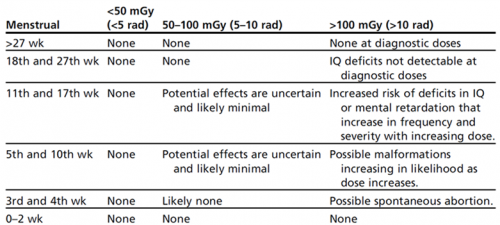
Everyone worries about imaging pregnant patients. As with most medical tests, it always boils down to risks vs benefits. What are the chances of causing mutations or cancers or a spontaneous abortion, and what is the risk of missing a critical injury? In general, reasonable studies involving a fetus at just about any point in gestation won’t cause major problems. At least as far as we know. What is not clear are the longer term, hard to measure effects. So the general philosophy should be to order just what you absolutely need, and shield the fetus during any studies other than of the abdomen/pelvis.

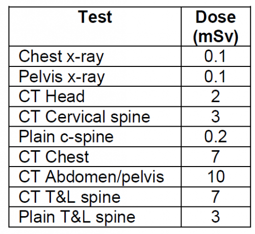
Now, to put these numbers into perspective, have a look at this list of delivered doses from common studies. The table above is listed in milliGrays, and this one is in milliSieverts. These are roughly comparable, except that the former is a measure of radiation dose absorbed, and the latter measures radiation delivered.

Bottom line: Think hard about the imaging you really need. If you generally do this for all patients, you probably won’t change your practice in pregnant women. Don’t worry about chest and pelvic x-rays. Shield the fetus for anything not involving the abdomen/pelvis. For major torso trauma, you probably will need CT of the chest/abdomen/pelvis. If so, do it right. Order with contrast so you don’t get substandard images that need to be repeated.

Print Friendly, PDF & Email

Trauma In Pregnancy 3: Tips & Tricks

Here is a list of practical tips for trauma professionals from prehospital and beyond:

  • Apply supplemental oxygen from the get-go, in the field if possible
  • Start an IV or IO and begin crystalloid infusion immediately
  • Bump the patient to her left. This is most easily done with the spine board in place. Just put a rolled up towel under the right side of the board.
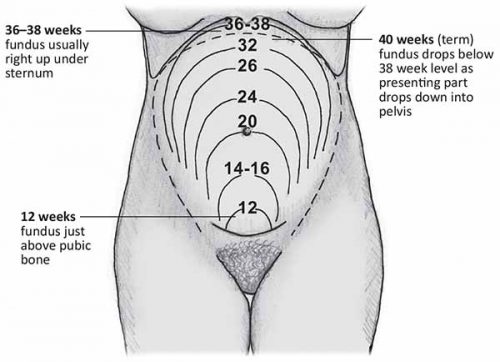
  • Quickly estimate how pregnant the patient is if she can’t tell you. Use the fundal height diagram below. Key measurements to remember:
    • Umbilicus = 20 weeks: entering the possible viability range if delivered emergently
    • Xiphoid = approaching full term

  • If the patient is 20 weeks+ pregnant, only transport to a trauma center (Level I or II).
  • At the hospital, reassess fundal height to more accurately estimate viability
  • If the fetus may be viable (23-24 weeks), call OB to the resuscitation area. But remember, they have nothing to offer until the trauma team secures the safety and stability of the mother. So don’t let them in the room until you have finished your entire evaluation.  Nothing they can do (e.g. monitor) is helpful during the acute phase of the resuscitation.
  • Once the mother is stabilized, assess fetal heart tones and apply monitors for fetal heart rate and maternal contractions.
  • Add coag studies to your lab panel. Fetal and placental injury can cause clotting problems.
  • Consider giving Rhogam in all major trauma cases if the mother is Rh negative
  • Obtain a KB test to look at the amount of fetal blood in the maternal circulation. Why do this if you are going to give Rhogam anyway? Answer: sometimes the amount of fetal blood leakage is greater than that covered by a single vial of Rhogam.
  • Plan your imaging intelligently. The next section covers this topic in more detail.
  • If needed, chest tubes should be inserted 1-2 intercostal spaces higher than usual.
  • The life-saving tetanus shot is safe in pregnant patients.


Print Friendly, PDF & Email