Category Archives: General

TraumaMedEd Newsletter for August

The newest TraumaMedEd newsletter is out! It was emailed to subscribers on Monday. This months focus is prehospital, with articles on the following:

  • Hare traction application
  • Trauma care and HIPAA, demystified
  • Trauma patient survival and air transport
  • What happens when you violate resuscitation guidelines for traumatic cardiac arrest?
  • Handoff issues between prehospital and the trauma team

Download the newsletter here!

Subscribe to the newsletter by clicking here!

Print Friendly, PDF & Email

Trauma Residents: How To Remember Liver Anatomy

In trauma surgery, operative management of liver injury is usually messy business, with little time for nice anatomic resections. However, an understanding of the basic anatomy, especially that of the vascular supply is crucial for saving your patient.

A cool tool for remembering Couinaud’s segments and the overall layout of liver anatomy was published in the Archives of Surgery recently. It makes use of a model, which consists of your hand! Just make a fist with your right hand and tuck the thumb behind the other fingers.


The fingers can then be numbered according to the Couinaud segments, with the caudate lobe (segment 1) represented by the thumb that is tucked away. The PIP joints represent the plane that the portal vein runs through, with branches going to upper and lower segments. Note how the ring finger normally lies a little more anterior than the little finger in this position, just like the sectors of the right lobe.

The creases between the fingers represent the left, middle and right hepatic veins.


The right hepatic vein is located between the right anterior and posterior sectors and the left hepatic vein sits between the left medial and lateral sectors. The middle hepatic vein is in between the left and right hemi-liver.

Bottom line: This “handy” liver model is available immediately in the OR and is already sterile. It can help visualize liver structures that may be injured quickly and accurately to speed your operative approach to the problem.

Reference: A Handy Tool to Teach Segmental Liver Anatomy to Surgical Trainees. Arch Surg 147(8):692-693, 2012.

Print Friendly, PDF & Email

PAs and NPs In Level I Trauma Centers

Trauma service staffing is important to maintaining trauma center status. Teaching centers in the US have been grappling with resident work hour rules, and non-teaching centers have always had to deal with how to adequately staff their trauma service. What is the impact of staffing a trauma center with midlevel practitioners (MLPs) such as physician assistants and nurse practitioners?

A state designated Level I trauma center in Pennsylvania retrospectively examined the effect of adding MLPs to an existing complement of residents on their trauma service. They examined the usual outcomes, including complications, lengths of stay, ED dwell times and mortality. 

Here are the more interesting results:

  • ED dwell time decreased for trauma activations and transfers in, but it increased for trauma consults. Of note, data on dwell times suffered from inconsistent charting.
  • ICU length of stay decreased significantly
  • Hospital length of stay decreased somewhat but did not achieve significance
  • The incidence of most complications stayed the same, but urinary tract infection decreased significantly
  • There was no change in mortality

Bottom line: There is a growing body of literature showing the benefits of employing midlevel providers in trauma programs. Whereas residents may have a variable interest in the trauma service based on their career goals, MLPs are professionally dedicated to this task. This study demonstrates a creative and safe solution for managing daily clinical activity on a busy trauma service.

Reference: Utilization of PAs and NPs at a level I trauma center: effects on outcomes. J Amer Acad Physician Assts, July 2011.

Print Friendly, PDF & Email

Shift Work And Fatigue In Air Medical Crews

Most trauma professionals are shift workers to one degree or another. It is well documented that sleep problems and fatigue can occur with this type of work, depending on the structure of the shift. A number of studies have been carried out in physicians and prehospital providers. But what about prehospital air crews?

Air medical providers are faced with two challenges: critically ill and injured patients and a challenging work environment. Typically, work consists of 12 or 24 hour shifts, and all of this is conducive to sleep problems and fatigue. 

The University of Pittsburgh looked at this problem, performing a battery of questionnaires and cognitive tests in their air medical service before and after each shift. They studied 37 subjects, and found the following interesting tidbits:

  • 95% of all crew members had poor baseline sleep quality
  • Fatigue levels decreased over the shift (both 12 and 24 hr)!
  • Crews were able to get some sleep while on duty (1 hour in a 12 hour shift, 7 hours in a 24 hour shift)
  • There was a mild increase in cognitive test performance at the end of the shift, although it was not statistically significant

Bottom line: Don’t anyone try to generalize these results to all flight crews! This was a sample of a single flight service, and is not necessarily representative of others. Poor baseline sleep quality is likely due to the fact that many flight nurses and paramedics hold other jobs. In this particular case, the decreasing fatigue may simply be due to the fact that they are encouraged to get some rest while on duty and actually do it. Make sure that your agency has fatigue reducing and fatigue avoidance policies and procedures. It’s for your safety as well as your patient’s!

Related posts:

Reference: The effect of shift length on fatigue and cognitive performance in air medical providers. Prehosp Emerg Care (early online, 2013)

Print Friendly, PDF & Email

How To Remove An Impaling Object

The books all say “transport the patient with an impaling object in place” and “only take the impaling object out in the operating room.” Is this realistic? How do you actually take that knife out?

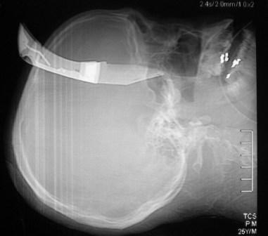
First, you need to decide if the patient belongs in the OR right now. Are they hemodynamically unstable? Is there obvious arterial bleeding? If so, don’t dawdle. Proceed to the operating room and surgically expose the problem completely.

If the patient is safe to stay in the ED, do what you need to figure out the exact anatomy of the wound (and object). This may involve imaging, usually CT scan. Once the exact position of the object is understood, build an anatomical picture of the situation in your mind. What named arteries might be involved? What other vital structures? 

Given this anatomic information, a decision can then be made regarding the best location for removal. The majority of the time, this will be in the operating room. It is best to obtain optimum surgical exposure prior to pulling it out. In the abdomen, this is easy. However, some areas (skull, sinuses) are tricky and may not require exposure of the end of the tract. Visualization of the remaining hole(s) is key so that bothersome bleeding can be recognized immediately.

The object should be grasped firmly and carefully and removed in one smooth motion. Visual monitoring for five minutes will virtually eliminate the presence of bleeding. If it does occur, then deeper exploration is warranted. In the awake patient, I generally push gently on either side of the entry point prior to and during the pull to provide some sensory distraction. Then I hold pressure on the site for 5 minutes (no peeking) to assure myself that there is no bleeding.

And don’t forget the forensics! Let the police photograph the patient. Handle the object carefully so as not to disturb any fingerprints. Place it carefully in a paper bag, labelled appropriately. And always make sure that a chain of evidence form is properly filled out so it and the object itself can be handed over to the proper authorities.

Print Friendly, PDF & Email