Category Archives: General

Management of CSF Otorrhea/Rhinorrhea

The management of CSF leaks after trauma remains somewhat controversial. The literature is sparse, and generally consists of observational studies. However, some general guidelines are supported by large numbers of retrospectively reviewed patients.

  • Ensure that the patient actually has a CSF leak. In most patients, this is obvious because they have clear fluid leaking from ear or nose that was not present preinjury. Here are the options when the diagnosis is less obvious (i.e. serosanguinous drainage):
    • High resolution images of the temporal bones and skull base. If an obvious breach is noted, especially if fluid is seen in the adjacent sinuses, then a CSF leak is extremely likely.
    • Glucose testing. CSF glucose is low compared to serum glucose.
    • Beta 2 transferrin assay. This marker is very specific to CSF. However, the test is expensive and results may take several days to a few weeks to receive. Most leaks will have closed before the results are available, making this a poor test.
  • Place the patient at bed rest with the head elevated. The basic concept is to decrease intracranial pressure, which in turn should decrease the rate of leakage. This same technique is used for management of mild ICP increases after head injury.
  • Consider prophylactic antibiotics carefully. The clinician must balance the likelihood of meningitis with the possibility of selecting resistant bacteria. If the likelihood of contamination is low and the patient is immunocompetent, antibiotics may not be needed.
  • Ear drops are probably not necessary. They may confuse the picture when gauging resolution of the CSF leak.
  • Wait. Most tramatic leaks will close spontaneously within 7-10 days. If it does not, a neurosurgeon or ENT surgeon should be consulted to consider surgical closure.

References:

  1. Brodie HA, Thompson TC. Management of complications from 820 temporal bone fractures. Am J Otol, 1997;18:188-197.
  2. Brodie HA. Prophylactic antibiotics for posttraumatic cerebrospinal fluid fistulas. Arch Otolaryngol Head, Neck Surg. 123:749-752.
Print Friendly, PDF & Email

Don’t Get S#!++y Xrays!

This has probably happened to you. You get a consult to see a trauma patient in your ED. As you walk into the room, you practically run into the massive portable x-ray machine sitting next to the patient. They’ve had some pretty significant blunt force to their leg, and the imaging has been ordered to rule out a fracture.

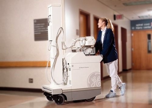
Sure, it’s convenient. The patient can stay right in the room. And it might be a little faster, especially if the regular x-ray department is a bit backed up. But let’s look at the my favorite indicator again, the juice to squeeze ratio

The control of the x-ray beam is not as good as with the fixed equipment. There just isn’t the same range of control available in the portable machines, which becomes important when nonstandard imaging and techniques are needed (think morbidly obese patient).

Placement of the x-ray plate can also be sub-optimal. This is especially true when biplanar images (i.e. AP and lateral) are requested, which is very common for fracture diagnosis. 

There may be additional exposure to radiation, especially to healthcare personnel or other patients. Someone has to hold that plate for the lateral image. Or other patients may be nearby, and shielding is not the same as in the rooms in the radiology department.

Bottom line: Sure, getting a portable x-ray may be the easy way to go. But only use if for studies that you absolutely must get quickly, and in which fine detail is not important. The standard chest and/or pelvis x-rays during trauma resuscitations fall into this category. But if you want the best quality imaging for diagnosis, and want to avoid repeat imaging, send your patient to the department to get some real images. Don’t settle for crappy ones!

Related posts:

Print Friendly, PDF & Email

March Trauma MedEd Newsletter is Available!

The March newsletter is now available! Click the image below or the link at the bottom to download. This month’s topic is Imaging.

In this issue you’ll find articles on:

  • CT contrast via IO catheter
  • FAST exams in children
  • CT scanning before transfer to pediatric trauma centers
  • Radiation exposure in pediatric trauma
  • IV contrast in trauma
  • And more!

Subscribers received the newsletter first over the weekend. If you want to subscribe (and download back issues), click here.

Download the newsletter here!

Print Friendly, PDF & Email