EAST 2014: The Flat Vena Cava Sign?

I’ve previously blogged about the flat vena cava sign as an indicator of low volume status in trauma patients. One of the papers at EAST takes another look at this tool, and had a surprisingly negative result.

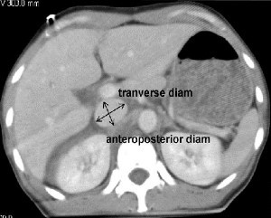
A retrospective study at George Washington University was carried out over a one year period. They looked at all of their highest level trauma activation patients who also underwent CT scan of the abdomen. Images were read by three radiologists and inter-rater reliability was reviewed. The transverse to anteroposterior diameter ratios were calculated to determine flatness.

Here are the factoids:

  • 276 patients met enrollment criteria, and were mostly male and blunt trauma
  • The IVC was nearly round in 21% of patients and collapsed in 26%
  • There was no association between IVC shape and shock index, blood pressure, Hbg, lactate, urgent operation, angiography or length of stay
  • There was also no association between IVC shape and blood transfusion or death
  • Correlation of the reads between radiologists was good

So what gives? A paper I reviewed three years ago in the Journal of Trauma came to a different conclusion. They found that a flat IVC on CT scan (defined as a transverse to AP ratio of 4:1 or greater) was associated with a significantly higher chance of receiving more crystalloid or blood, as well as requiring an operation within 24 hours. 

First, the newer work is an abstract, so a lot may be unsaid at this time. This is why I encourage everyone to always read the entire paper! The published paper involved a smaller series (114 patients), but it was prospective and had reasonable statistical analyses.

Bottom line: This is a presentation that I’ll have to sit through and ask the authors why they didn’t find the same results as the older paper. For now, continue to use the flat IVC sign as a potential sign of trouble ahead. I’ll report more on this one later in January.

Reference:

  • Inferior vena cava size is not associated with shock following injury. EAST 2014, oral paper 12.

Related post:

The Best (And Worst) Of EAST

The EAST meeting is upon us in just a few weeks! I’ll be attending (and tweeting) during this annual event. But in the meantime, I’d like to provide some commentary on some of the best and worst abstracts that will be presented.

Starting tomorrow, I’ll summarize one interesting paper/poster per day. I’ll also provide some perspective as to why it looks important (or not). Remember, my impressions are based on just an abstract. There is a lot that is left unsaid in the preparation of these brief research summaries. Sometimes, those things are crucial and totally destroy the work promised in the abstract.

I’ll also be listening to the presentations of the papers I critique at the meeting so I can get the real story, and I’ll share those with you as well next month.

What Would You Do? Final Answer!

Lots of great comments about this case yesterday. So you’re looking for reasons why this 8 year old child is having blood pressure problems while undergoing craniotomy. On first pass through the ABCs, everything looks good. Looks. That’s not enough. 

The airway is definitely good. It’s been physically checked. But what about breathing? Lung sounds seem to be good, but in children they can be transmitted from the other side. There are a few solutions. If you have fluoro or quick access to a chest xray, that could be used. However, quickness is the key. If in doubt and the pressure is really becoming a problem, consider empiric needle decompression or finger thoracostomy if you want to avoid chasing with a chest tube on each side.

What about circulation? We’ve ruled out just about every source of bleeding but the abdomen. No FAST? Then why not fall back on DPL? Sure, it’s been a while since you’ve done it. And probably even longer since you’ve done one on a child.

DPL tips for kids: You’re really interested in the tap, not the lavage. If there’s enough bleeding in the abdomen to cause hypotension, you’ll find it quickly. Use open technique (not Seldinger) and insert the catheter far enough to cover all the tiny holes. One reader suggested using a suction cathether (like a Poole sucker). The only issue is one of control so the amount of blood removed can be measured. (Bonus tip: if you ever do the lavage part in a child, use 10cc saline or lactated ringers per kg).

In this case, the chest was negative, and there was no blood in the abdomen. The culprit was D (disability in ATLS jargon). And unfortunately, hypotension from CNS causes is universally an indicator of impending doom.

What Would You Do? Part 2

Yesterday, I wrote about an 8 year old bicyclist struck by a car. He had sustained a head injury with a large epidural hematoma and was taken immediately to the OR for evacuation. During the case, the vitals began to suffer. The question was, what would you do as you are called into the room to “fix” this?

There was some excellent chatter on this, and universally, people voted to stick to the ABCs. As taught in ATLS, when the vital signs go awry, the only problems that can cause this degree of sudden derangement start with A, B, C and occasionally D.

The first thing to do is recheck the adequacy of the airway. Sure, the child is intubated. But the neurosurgeon had to position the head, and anesthesia’s access to this area is more limited than in other procedures. Your anesthesiologist is able to put a fiberoptic scope into the mouth and visualize that the tube does indeed pass through the vocal cords. Oxygen saturation is 100% and ETCO2 is 33.

Next, check breathing. The numbers above are helpful, but not the complete answer. Get access to the chest, look and listen. There is good, symmetric chest rise with ventilation, and breath sounds are completely normal bilaterally. The trachea is midline.

Now, is there any circulation problem (bleeding)? There are 5 areas where you can bleed to death:

  • Chest – the initial chest xray and your exam make this unlikely.
  • Fractures – you examine all extremities and can’t find any gross fractures
  • Pelvis – the initial portable xray did not show any fractures, and your exam is normal
  • Floor – your shoes are not getting sticky and the OR table is clean (except for the neurosurgeon’s mess)
  • Abdomen – ???

Hmm, the abdomen appears a bit distended, and it’s a bit tympanitic. This is a problem area! Let’s say FAST is not available to you, and the pressure is not improving with fluid and pressors.

How can you evaluate this child’s abdomen in this situation? I want the gory details, and will provide answers tomorrow.

What Would You Do?

Here’s an interesting clinical case. How would handle this difficult situation?

An 8 year old boy is struck by a car while riding his bicycle. He has obvious head trauma, and medics quickly transport him to your trauma center. He is comatose and posturing with a GCS of 5 (E1 V1 M3). RSI and intubation is carried out, and a full exam is done. Only head trauma is noted, with the right pupil a few millimeters larger than the left. The remainder of the physical exam is unremarkable, with the exception of a few extremity abrasions. FAST is negative, as are portable chest and pelvis xrays.

The child is quickly transported to CT, and this is discovered:

He is immediately transported to the OR, where your pediatric neurosurgeon immediately begins a craniotomy to evacuate the epidural hematoma. 

Thirty minutes into the case, you are summoned to the OR because the patient’s blood pressure is dropping and is not responding as expected to fluids and a touch of pressor.

What do you need to think about, and what would you do?

Please comment or tweet all the details. Answers tomorrow!

Home of the Trauma Professional's Blog

Do you want to get a daily email every time there’s a new post? See what I’m up to.

Click here to get details and subscribe!

[accua-form fid=”1″]

[mc4wp_form id=”2023″]