All posts by The Trauma Pro

5 Guidelines For Diaphragmatic Injury

Today’s post is another review of some of the practice guidelines published by the Eastern Association for the Surgery of Trauma (EAST).  This one covers the evaluation and management of diaphragmatic injury.

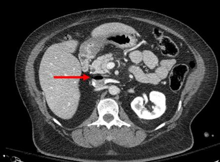
Diaphragm injury is a troublesome one to diagnose. It is essentially an elliptical sheet of muscle that is doubly-curved, so it does not lend itself well to diagnosis by axial imaging. Addition of sagittal and coronal reconstructions to a thoraco-abdominal CT has been helpful, but still has a far from perfect diagnostic record.

From an evaluation standpoint, there are several possibilities:

  • Observation – not generally recommended. It is usually combined with imaging such as chest x-ray to see if interval changes occur that would indicate the injury.
  • Chest x-ray – this is not often diagnostic, but when herniation of abdominal contents is obvious the patient most assuredly has an operative problem.
  • Thoraco-abdominal CT scan – this technology keeps getting better, especially with thinner cuts and different planes of reconstruction. Sometimes even subtle injuries can be detected. But this exam is still imperfect.
  • Laparoscopy or thoracoscopy – this technique yields excellent accuracy when the injury is in an area that can be viewed from the operative entry point chosen.
  • Laparotomy or thoracotomy – this is the ultimate choice and should be nearly 100% accurate. It is almost the most invasive and has more potential associated complications.

EAST reviewed a large body of literature and selected 56 pertinent papers for their quality and design. They critically reviewed them and applied a standard methodology to answer several questions.

Here are the questions with the recommendations from EAST, along with my comments:

  • Should laparoscopy or CT be used to evaluate left-sided thoraco-abdominal stab wounds? First, these patients must be hemodynamically stable and not have peritonitis. If either is present, there is no further need for diagnosis; a therapeutic procedure must be performed.
    Left sided diaphragm injuries from stabs are evil. The hole is small, and since the pressure within the abdomen is greater that the chest, things always try to wiggle their way through this small hole. It can remain asymptomatic if the wiggler is just a piece of fat, but can be catastrophic if a bit of the stomach or colon pushes through and becomes strangulated. Furthermore, these holes enlarge over time, so more and more stuff can push up into the chest.
    EAST recommends the use of laparoscopy for evaluation to decrease the incidence of missed injury. However, if the injury is in a less accessible location (posterior), the patient has body habitus issues, or adhesions from previous surgery may lead to incomplete evaluation, laparotomy should be strongly considered.
  • Should operative or nonoperative management be used to evaluate right-sided thoraco-abdominal penetrating wounds? Note that this is different than the last question. All penetrating injuries are included (stabs and gunshots), and this one is for management, not evaluation. And the same caveats regarding hemodynamic stability and peritonitis apply. It applies to both stabs and gunshots.
    Unlike left-sided injuries, right-sided ones are much more benign. The liver keeps anything from pushing up through small holes, and they do not tend to enlarge over time due to this protection. For that reason, EAST recommends nonoperative management to reduce mortality and morbidity related to operation.
  • Should stable patients with acute diaphragm injury undergo repair via an abdominal or thoracic approach? This question applies to any diaphragm injury that requires operation, such as right-sided penetrating injury or any blunt injury. EAST recommends an approach from the abdomen to reduce morbidity and mortality. Since abdominal injury frequently occurs in these cases, an approach from the chest limits the ability to identify and repair abdominal injuries. Otherwise, you may find yourself doing a laparotomy in addition to the thoracotomy.
  • Should patients with delayed visceral herniation through a diaphragm injury undergo repair via an abdominal or thoracic approach?  For years, the preferred approach for delayed presentations has been through the chest because the injury is easier to appreciate and repair.  However, if ischemic or gangrenous viscera are present, it will be more difficult to manage and repair from the chest. EAST does not make a specific recommendation for this question and suggests the surgical approach be determined on a case by case basis.
  • Should patients with an acute diaphragm injury from penetrating injury without concern for other intra-abdominal injuries undergo open or laparoscopic repair? The quality and quantity of data addressing this question were very low, but EAST recommends laparoscopy for repair of these injuries to reduce morbidity and mortality. This includes blunt injuries, which tend to be larger. There were some conversions to an open procedure, especially in the blunt cases. The usual caveats on exposure, injury location, body habitus and previous surgery apply.

Reference: Evaluation and management of traumatic diaphragmatic injuries: A Practice Management Guideline from the Eastern Association for the Surgery of Trauma. J Trauma 85(1):198-207.

4 Guidelines For the Management Of Bladder Injury

The Eastern Association for the Surgery of Trauma (EAST) has been at the forefront of trauma practice guideline dissemination for decades. They recently published a set of recommendations for managing patients with bladder injury. These injuries are not commonly encountered by trauma professionals, and I thought a refresher on current thinking on their management was in order.

Using the usual methodology, the trauma literature was scanned for papers dealing with this topic. After screening for quality, the field was narrowed to 17 papers which were used to formulate the published recommendations. These cover imaging and management questions that frequently come up during the evaluation of these patients.

Following are the questions raised, the EAST recommendations, and my commentary about them:

    • In patients with abdominal / pelvic trauma, should retrograde CT cystography vs no imaging be used to diagnose a bladder injury? This seems like a silly question, but the answer lies in the details. It all boils down to the likelihood of injury. And how does one determine likelihood? By looking at the urine and the fracture patterns around the bladder. Patients with microscopic hematuria are very unlikely to have a bladder injury, and any type of bladder imaging in these patients (cystogram, CT cystogram) is almost never positive, and so is not indicated. This is the reason that ordering a urinalysis in major trauma patients is not recommended. However, if gross hematuria is present, CT cystogram is recommended. The sensitivity and specificity are nearly perfect. Just be sure to do a true cystogram by actively filling the bladder with contrast via a urinary catheter. Passive filling of the bladder with urine from the IV contrast misses about half of all the injuries. Also, strongly consider adding CT cystogram in patients with widening of the pubic symphysis. This injury pattern is frequently associated with bladder injury.
    • In patients with intraperitoneal bladder rupture from blunt trauma, should operative or nonoperative management be used to decrease complications? Another silly question? In general, intraperitoneal bladder ruptures do not heal on their own, so urine continues to bathe the peritoneal cavity until the injury is fixed. The review article recommended that operative repair be performed in all of these cases. 
    • In patients with extraperitoneal bladder rupture from blunt trauma, should operative or nonoperative management be used to decrease complications? Patients with a simple extraperitoneal bladder injury should undergo nonoperative management. These injuries usually heal and seal within about 10 days. However, patients with this type of bladder injury that is more complicated (bone spicules piercing the bladder, concomitant vaginal or rectal injury, bladder neck injury) should undergo operative repair in order to decrease the complication rate. One additional group that should be repaired: patients with pubic diastasis that will require operative fixation. The bladder should be repaired at the time of the orthopedic procedure to avoid bathing the new hardware in urine.
    • In patients who have undergone operative or nonoperative management of bladder injury, should bladder closure be assessed with cystogram or not? This one depends on the type and complexity of injury. For simple intraperitoneal bladder injuries that were operatively repaired, no followup cystogram is required. More complex repairs should be evaluated by cystogram before removing the urinary catheter. Finally, simple extraperitoneal injuries should also have a cystogram obtained before removing the catheter. My magic number for obtaining followup studies is 10 days. There is no real science behind this, and no one has systematically looked at 5 vs 7 vs 10 vs 14 days. This one is based only on personal experience.

And by the way, most simple bladder injuries (both intra- and extra-peritoneal) can be easily repaired using two layers by your friendly neighborhood trauma surgeon. More complex injuries are generally best left to the urologist.

Reference: Management of blunt force bladder injuries: A practice management guideline from the Eastern Association for the Surgery of Trauma. J Trauma 86(2):326-336, 2019.

Coming Soon! New Site For Trauma PI!

One of the most common requests I get is to provide more detailed content on Trauma Performance Improvement! To that end I am putting together a collection of print and video content on a new website that will address the things you really want to hear about but can’t find anywhere else.

Here’s a sample listing of some of the topics that will be covered:

  • Writing a good PI plan
  • Loop closure – basic to advanced
  • Involving your TMD
  • PRQ preparation
  • Creating workable practice guidelines
  • Crafting a Massive Transfusion Protocol that works for you
  • How to calculate your optimal number of trauma registrars
  • Preparing for your site survey
  • How to read your TQIP report
  • What is OPPE and how do I do it?
  • Integrating PI with your registry
  • How to interpret the Orange Book

If you want to be one of the first to get access to this content, please fill out the form by clicking here. Your name will be placed on my early bird e-mail list. I’ll provide regular updates on the opening date, and solicit your ideas on specific content you would like to see.

Subscribe to the mailing list now!

Air Embolism From an Intraosseous (IO) Line

Intraosseous (IO) lines are a godsend when we are faced with a patient who desperately needs access but has no veins. The tibia is generally easy to locate and the landmarks for insertion are straightforward. They are so easy to insert and use, we sometimes “set it and forget it”, in the words of infomercial guru Ron Popeil.

But complications are possible. The most common is an insertion “miss”, where the fluid then infuses into the knee joint or soft tissues of the leg. Problems can also arise when the tibia is fractured, leading to leakage into the soft tissues. Infection is extremely rare.

This photo shows the inferior vena cava of a patient with bilateral IO line insertions (black bubble at the top of the round IVC).

During transport, one line was inadvertently disconnected and probably entrained some air. There was no adverse clinical effect, but if the problem is not recognized and the line is not closed properly, there could be.

Bottom line: Treat an IO line as carefully as you would a regular IV. You can give anything through it that can be given via a regular IV: crystalloid, blood, drugs. And even air, so be careful!

Giving TXA Via An Intraosseous Line?

Seriously injured patients frequently develop coagulopathy, which makes resuscitation (and survival) more challenging. A few years ago, the CRASH-2 study lent support for using tranexamic acid (TXA) in select trauma patients to improve survival. This drug is cheap and has antifibrinolytic properties that may be beneficial if given for life-threatening bleeding within 3 hours of initial injury. It’s typically given as a rapid IV infusion, followed by a slower followup infusion. The US military has adopted its routine use at forward combat hospitals.

But what if you don’t have IV access? This can and does occur with military type injuries. Surgeons at Madigan Army Medical Center in Washington state tried using a common alternative access device, the intraosseous needle, to see if the results were equivalent. This study used an adult swine model with hemorrhage and aortic crossclamping to simulate military injury and resuscitation. Half of the animals then received IV TXA, the other half had it administered via IO. Only the bolus dose was given. Serum TXA levels were monitored, and serial ROTEM determinations were performed to evaluate coagulopathy.

Here are the factoids:

  • The serum TXA peak and taper curves were similar. The IV peak was higher than IO and approached statistical significance (0.053)
  • ROTEM showed that the animals were significantly hyperfibrinolytic after injury, but rapidly corrected after administration of TXA. Results were the same for both IV and IO groups.

Bottom line: This was a very simple and elegant study. The usual animal study issues come into play (small numbers, pigs are not people). But it would be nearly impossible to have such a study approved in humans. Even though the peak TXA concentration via IO is (nearly significantly) lower, this doesn’t appear to matter. The anti-fibrinolytic effect was very similar according to ROTEM analysis.

From a practical standpoint, I’m not recommending that we start giving TXA via IO in civilian practice. We don’t typically see military style injuries, and are usually able to establish some type of IV access within a reasonably short period of time. But for our military colleagues, this could be a very valuable tool!

Reference: No intravenous access, no problem: Intraosseous administration of tranexamic acid is as effective as intravenous in a porcine hemorrhage model. J Trauma 84(2):379-385, 2018.