All posts by The Trauma Pro

Best of EAST 2024 #1: MAP And Spinal Cord Injury

The use of elevated mean arterial pressure (MAP) to help manage spinal cord injury has been a mainstay of treatment for years. The concept is similar to that used for management of severe traumatic brain injury. The theory is that there may be areas of the brain that are damaged, but not irretrievably so. Increasing MAP should improve perfusion and may protect areas in jeopardy from secondary injury.

As with so much in neurotrauma, few large and/or prospective studies exist. Although most centers have specific algorithms and MAP goals, optimal treatment still needs to be determined.

EAST sponsored a prospective, multicenter study to identify factors influencing neurologic outcomes after spinal cord injury. MAP augmentation was monitored, specifically its impact on the American Spinal Injury Association (ASIA) score between admission and discharge.

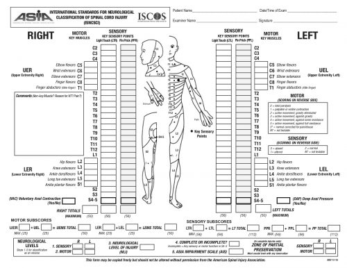
The ASIA Score is calculated by performing a very detailed exam consisting of myotomal motor function, a dermatomal sensory exam, and an anorectal exam. The exam takes quite some time to complete. The copy of the worksheet below should give you an idea of the level of detail:

The study was performed over 20 months, and 19 centers participated. They entered 222 patients, but only 164 had pre- and post-ASIA scores for comparison.

Here are the factoids:

  • Of the 164 patients studied, only 36 improved vs. 128 that showed no improvement by ASIA score
  • Demographics, hospital and ICU length of stay, and mortality were not significantly different between the groups
  • ISS was nearly identical (23 vs 25)
  • Three-quarters of injuries were to the cervical spine, about 10% to the lumbar spine, and the remainder to the thoracic spine. There was no correlation between injury location and recovery.
  • Presentation in the trauma bay (blood pressure, pulse, MAP, lactate, and Hgb) were the same in both groups
  • The MAP goal of >85 mm Hg was met about 75% of the time in both groups
  • Duration of MAP therapy was the same for the two groups, from 99-113 hours
  • There was a trend toward increased cardiac issues (atrial fibrillation, v-tach, elevated troponin) in the group with improved spinal cord recovery. This may be due to the medications used to increase MAP.

Bottom line: This is very interesting work and will make us question the utility of MAP therapy for spinal cord injury. However, this is not a cut-and-dried conclusion. Here are several things that come to mind:

  • What was the definition of “improvement?” ASIA is a complicated scoring system with many steps in the evaluation. Usually, the results are condensed into an overall “ASIA Impairment Scale,” or AIS.
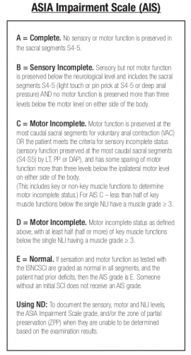
    The AIS is not very granular, meaning that each step in the scale represents a large difference in function. Could patients have had improvements that did not change the AIS score but were functionally significant for the patient? For example, an improvement from a C5 to a C6 level makes a big difference in daily activities.
  • Was the study large enough? It is difficult to accumulate a large series of spinal cord injury patients. Combining this point with the previous one, was the statistical power present even to detect a meaningful difference in the AIS?
  • Was MAP>85 torr maintained reliably and for long enough? Patients had MAP therapy for just over four days, and it was only maintained above the threshold about 75% of the time. We have good evidence in the brain injury literature that a single bout of hypotension in patients with severe TBI significantly increases mortality. Could it be that maintaining increased spinal cord perfusion is equally important? Could a single low MAP cause damage? It would be interesting to see if patients who had very consistent MAP therapy, say greater than 90% or 95% of the time, had any difference in outcomes. Unfortunately, I suspect that the numbers would be far too low to prove anything.

This abstract brings up some interesting questions. However, I would not consider throwing out the use of MAP goals based on it. We need more patients to study and be better at applying this treatment if we hope to uncover whether it really works.

Reference: Does mean arterial pressure augmentation improve neurological recovery of blunt spinal cord injuries: an EAST multicenter trial. EAST 2024 Podium paper #1.

Coming Soon! The Best Of EAST 2024

The 37th Annual Assembly of the Eastern Association for the Surgery of Trauma is just around the corner! And, as in previous years, I will be publishing regular posts on some of the abstracts I find the most interesting. Here are some of the topics I’ve selected:

  • MAP and spinal cord injury
  • VTE in pediatric patients
  • Chest irrigation and retained hemothorax
  • Accuracy of eFAST
  • More on the 35mm rule for pneumothorax
  • Pan-scanning and missed injuries
  • King Airway vs i-Gel Airway
  • Whole blood transfusion in pediatric patients
  • Whole blood and VTE risk
  • VTE prophylaxis in patients undergoing acute neurosurgical intervention

For each abstract covered, I will present the findings and give a short critique. Finally, I will provide some questions for the authors to consider, as they very same ones could come from the audience at their presentation!

If you have any particular abstracts you would like me to cover, please list in the comments section below and I will get it on the list!

Should I Apply Compression Devices To Patients With DVT?

Everyone knows that venous thromboembolism (VTE) is a potential problem in hospitalized patients, and especially so in trauma patients. Several groups of them are at higher risk by virtue of the particular injuries they have sustained and the activity restriction caused.

Nearly every trauma program uses some form of screening and prophylaxis in an attempt to reduce the occurrence of this problem, which can result in deep venous thrombosis (DVT) and/or pulmonary embolism (PE). Screening looks at patient factors such as age, obesity, and previous VTE, as well as injury risk factors like spine and pelvic fractures and decreased mobility.

Based on the screening protocol, prophylaxis may be prescribed depending upon the level of VTE risk, which is then balanced with bleeding risk from the brain, solid organ, or other injuries. The choices we have are primarily mechanical vs chemical and consist of compression devices (sequential or not) and various heparins.

But an age-old question continues to resurface: if a patient breaks through their prophylaxis and develops DVT, is it safe to apply compression devices to the extremity?

There has always been the fear that doing things that increase flow in the affected extremity may cause clots to dislodge and ultimately cause a PE. Seems logical, right? But we know that often, our common sense about things is completely wrong.  Couldn’t just moving around cause pieces to break off? A meta-analysis of 13 studies published in 2015 showed that early ambulation was not associated with a higher incidence of new PE. Furthermore, patients who suffered from pain in the affected extremity noted significant improvements with early ambulation.

If ambulation makes the pain better, could the veins be recanalizing more quickly? Another study examined a small group of 72 people with DVT receiving anticoagulants, half of whom were prescribed exercise and compression stockings and the other half stockings only. There was a huge amount of variability in the rates of recanalization, but ultimately, there were no significant differences with or without exercise.

So just lying in bed is not good, and exercise/ambulation may actually make people feel better. But interestingly, bedrest alone does not appear to increase the likelihood of PE! It does decrease the risk of developing problems other than the VTE, like pulmonary complications.

But what about compression devices? Common sense would say that you are intermittently  increasing pressures in the leg veins, which could dislodge any loose clots and send them flying to the lungs, right?

Unfortunately, I couldn’t find a paper from anyone who had the courage to try this. Or perhaps no institutional review board (IRB) would approve it. But the key fact is that every compression device manufacturer includes existing DVT as a contraindication in their product documentation. They don’t have any literature either, so I assume it’s an attempt to limit litigation, just in case.

Bottom line: Walking provides at least as much muscle compression as compression devices. But the simple truth is that we have no solid research that either supports or condemns the use of active compression devices in patients with known DVT. And we probably won’t, ever.

Compression stockings seem to be safe, but they really don’t do much. They are white, but don’t do much more than contribute to hospital clothing fashion. Since the manufacturers define existing DVT as a contraindication, application of their product would be considered an off-label use. So it looks like we cannot in good faith use these devices in patients with diagnosed DVT.

References:

  • Bed Rest versus Early Ambulation with Standard Anticoagulation in The Management of Deep Vein Thrombosis: A Meta-Analysis. PLOS One , April 10, 2015, https://doi.org/10.1371/journal.pone.0121388
  • Bed Rest or Ambulation in the Initial Treatment of Patients With Acute Deep Vein Thrombosis or Pulmonary Embolism: Findings From the RIETE Registry. Chest 127(5):1631-1636, 2005.
  • Does supervised exercise after deep venous thrombosis improve recanalization of occluded vein segments? A randomized study. J Thrombosis Thrombolysis 23:25-30, 2006.

Use Of Radio-opaque Markers In Penetrating Trauma

As I was browsing through my journal list this week, I ran into an interesting title for an article that is currently in press.

“The use of radio-opaque markers is medical dogma”

Catchy, especially since I love writing about dogma vs what is really supported by the literature. The author questions the justification of this practice and posits that there are risks to extrapolating information based on radiographs with markers placed by the trauma team.

OLYMPUS DIGITAL CAMERA

The author first reviewed the literature on the use of markers for penetrating injury, which started only recently, in 2002. Markers were initially used to precisely locate the penetration site since skin wounds (obviously) don’t show up on X-rays. Typically, these were just plain old paper clips. Some trauma professionals placed them directly over the wound. Others un-bent them and fashioned them into shapes that pointed to the exact location of the wound.

With the growing usage of CT scans to evaluate stable patients, modifications to the marker were made. Small arrow markers designed for use on x-rays were frequently used. However, even the very small ones could cause enough scatter on a CT scan to interfere with diagnosis. At some centers, Vitamin E capsules were taped on top of the wound. But thankfully, there are now special markers that can pinpoint the wound without degrading the tomographic image.

The author goes on to describe how gunshot wounds specifically are difficult to assess with a marker. Although the exact surface location may be noted, the underlying injuries vary due to size, distance, velocity, and trajectory change from tissue density or bone strikes. He also notes that it may not be wise to place a marker into a bloody field in a potentially combative patients.

The article concludes that the use of this technique for identifying anything other than surface location of penetrations lacks clinical evidence and is based only on expert opinion. Which essentially makes it dogma.

Bottom line: Here are my thoughts. First, the use of markers on penetrating wounds has been going on for much longer than the 20 years found in the trauma literature reviewed here. It has been a common practice among trauma surgeons for many, many decades. Most “seasoned” (old) trauma surgeons have been doing and teaching this for their entire careers. 

I concur that we have techniques like CT scan available to us now that provide an excellent view of the penetration trajectory. The skin wound is usually apparent on the scan, but may be improved with the use of a CT-approved marker.

So why still do this for the patient arriving in your trauma bay? An experienced trauma surgeon can get a good sense of the trajectory based on the entry point, the exit wound, and the location of any retained bullet or fragments. Rapid placement of some kind of marker on all wounds followed by a quick image allows them to roughly predict what was hit, and assess the possibility that there might be bleeding that would drive the team straight to the operating room. It can help direct the surgical exploration if imaging was unnecessary or contraindicated. 

So yes, this is dogma. The reality is that no one will ever be able to design a study that definitively evaluates the very soft outcomes that result from using this technique. But every senior trauma surgeon can easily cite numerous examples in their career when this method has been extremely useful. The lack of a study only means that there will never be any evidence-based guideline for the use of this technique. However, it is still acceptable to have a protocol based on substantial clinical experience. But as with all dogma, once that definitive study finally does comes along, the protocol must be modified to adhere to the findings of the study.

For now, keep using those markers! And I’m very interested in comments from both old and young trauma professionals on this topic.

Reference: The Use of Radio-opaque Markers is Medical Dogma, doi:10.1111/acem.1485, Dec 2023.

Liquid Plasma vs FFP: Impact On Your Massive Transfusion Protocol

In my last post, I discussed the growing number of choices for plasma replacement. Today I’ll look at some work that was done that tried to determine if any one of them is better than the others when used for the massive transfusion protocol (MTP).

As noted last time, fresh frozen plasma (frozen within 8 hours, FFP) and frozen plasma (frozen within 24 hours, FP) have a shelf life of 5 days once thawed. Liquid plasma (never frozen, LQP) is good for the 21 days after the original unit was donated, plus the same 5 days, for a total of 26 days.

LQP is not used at most US trauma centers. It is more commonly used in Europe, and a study there suggested that the use of thawed plasma increased short term mortality when compared to liquid plasma. To look at this phenomenon more closely, a group from UTHSC Houston and LSU measured hemostatic profiles on both types of plasma at varying times during their useful life.

All products were analyzed with thromboelastography (TEG) and thrombogram, and platelet count and microparticles, clotting factors, and natural coagulation inhibitors were measured. They chose 10 units of thawed FFP and 10 units of LQP, and assayed them every 5 days during their useful shelf life.

Here are the factoids:

  • Platelet counts were much higher in day 0 LQP (75K) vs day 0 thawed plasma (7.5K). Even at end of shelf life, the LQP was 1.5x higher than thawed (15K vs 10K).
  • Thrombogram showed that LQP had higher endogenous thrombin production until end of shelf life
  • TEG demonstrated that LQP had a higher capacity to clot that gradually declined over time. It became similar to thawed plasma at the end of its shelf life.
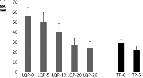
                         (TEG MA for liquid (LQP) and thawed (TP) plasma
  • Most clotting factors remained stable in LQP, with the exception of Factors V and VIII, which slowly declined

Bottom line: Liquid plasma sounds like good stuff, right? Although there are a few flaws in the collection aspect of this study, it gives good evidence that never frozen plasma has better coagulation properties when compared to thawed plasma. Will this translate into better survival when used in the MTP for trauma? One would think so, but you never really know until you try it. Our hospital blood bank infrastructure isn’t prepared to handle this product yet, for the most part. What we really need is a study that shows the survival advantage when using liquid plasma compared to thawed. But don’t hold your breath. It will take a large number of patients and some fancy statistical analysis to demonstrate this. I think we’ll have to look to our military colleagues to pull this one off!

Reference: Better hemostatic profiles of never-frozen liquid plasma compared with thawed fresh frozen plasma. J Trauma 74(1):84-91, 2013.