All posts by The Trauma Pro

What Is The Safest Extrication Method From A Car Crash?

Today’s post is directed to all those prehospital trauma professionals out there.

Car crashes account for a huge number of injuries world-wide. About 40% of people involved in them are initially trapped in the vehicle. And unfortunately, entrapped individuals are much more likely to die.

There are four basic groups (and their category in parentheses) of trapped car occupants:

  • those who can self-extricate or extricate with minimal assistance (self-extrication)
  • individuals who cannot self-extricate due to pain or their psychological response to the event, but can extricate with assistance (assisted extrication)
  • people who are advised or choose not to self-extricate due to concern for exacerbating an injury, primarily spine (medically trapped)
  • those who are physically trapped by the wreckage who require disentanglement (disentanglement and rescue)

Prehospital providers have several choices to help extricate patients  in the second and third categories: encourage self-extrication, rapid extrication without the use of tools, or traditional extrication where the vehicle is cut away to allow egress. The fourth category always requires tools for extrication.

Although rescue services try to minimize or mitigate unnecessary movement of the patient, stuff happens. Large and forceful movement is considered high risk, but smaller movement do occur. This is of particular concern in patients who might have a spine injury.

There have been a number of recent papers suggesting there might be greater benefits to self-extrication. A group of authors in the UK and South Africa designed a biomechanical study to test these methods of extrication in healthy volunteers.

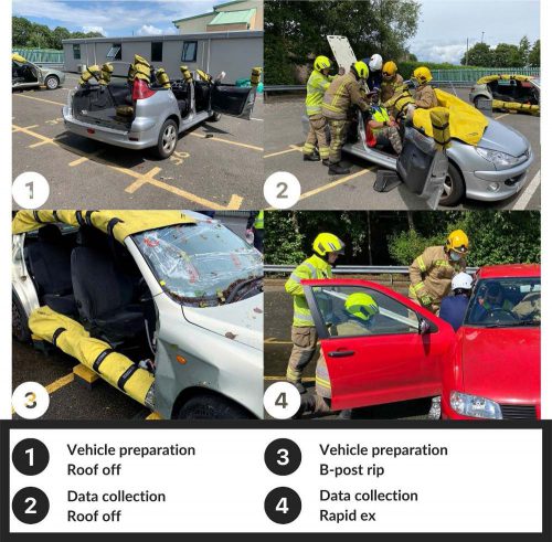
The authors wanted to find out exactly how much movement occurred using the various extrication techniques. The volunteers were fitted with an Inertial Measurement Unit, which measures the orientation of the head, neck, torso, and sacrum in real time.  The IMU can detect even very small changes in orientation of the body. The volunteers were placed in a standard 5-door hatchback sedans that were prepared for each type of extrication as seen above.

Here are the factoids:

  • A total of 230 extrications were performed for analysis
  • The smallest amount of maximal and total movement of body segments was seen in the self-extrication group
  • The greatest amount of movement was found in the rapid extrication group, with 4x to 5x the movement in the self-extrication group
  • The difference in body movement between the self-extrication group and all others was significant
  • In general, movement increased as extrication techniques progressed from roof removal to B post removal to rapid extrication

The authors concluded that self-extrication resulted in the smallest amount of movement and the fastest extrication time, and that it should be the preferred technique.

Bottom line: This is the first study that specifically evaluated spinal movement occurring with commonly used extrication techniques. Other similar studies have used a variety of measurement techniques, none of which are as precise as this. One potential weakness with this one is that it used healthy volunteers. But obviously, it is not practical to attempt anything like this with real, injured patients. 

Since we know that patients trapped in cars are more likely to die, time is of the essence. This study shows that self-extrication is both fast and safe with respect to spinal movement. The information will assist our prehospital colleagues in making the best decisions possible when faced with patients trapped in their car.

Reference: Assessing spinal movement during four extrication methods: a biomechanical study using healthy volunteers. Scand J Trauma  open access 30: article 7, 2022.

For PI Fans: Cribari, NFTI, And STAT!

I’ve published a two-part series on the Cribari matrix, Need For Trauma Intervention (NFTI), and the Standardized Triage Assessment Tool (STAT). These are performance improvement topics for the real nerds out there and can be found only on my Trauma PI website, TraumaMedEd.com.

If you are interested in optimizing trauma triage and trauma activations at your center, check out my posts by clicking this link:

https://www.traumameded.com/blog/

Optimizing Feedback To Referring Hospitals

The American College of Surgeons requires that referring hospitals provide feedback to prehospital providers and referring hospitals regarding the transfer process.

Failure to do so can actually result in a weakness or deficiency during a site visit. (Psst! Pay attention, referring hospitals if you want to start getting feedback. Read that first sentence again.) Sometimes the feedback is verbal, either in person or by phone. Many receiving centers send written letters outlining care and care issues. But unfortunately, some don’t do it at all, or only very inconsistently.

Harborview Hospital in Seattle is a very busy Level I center, with nearly 6,000 trauma admissions per year. More than half of their patients come from a huge catchment area including Washington state, Wyoming, Alaska, Idaho, and Montana. The amount of work to provide proper feedback on over 3,000 patients annually can be overwhelming.

They implemented a “U-link” program that provided access to patient chart info for the hospital sending each patient. It was HIPAA compliant, and login information was sent within 72 hours of patient arrival.

Here are the factoids:

  • 90 referring hospitals set up the U-link system
  • Care transcripts, radiology reports, and discharge summaries were the most frequently viewed items
  • The most desired feedback was on over- or under-resuscitation (89%), injuries (84%), appropriateness of transfer (78%), and deviation from ATLS protocols (76%)
  • Information was used for education (100%), systems analysis (99%), and performance improvement (PI, 92%)

Bottom line: Your referral partners crave feedback on the patients they send! Develop a system that guarantees it on each patient at a reasonable time after admission. You may or may not be able to link them into your specific electronic medical record, but you can certainly send out informational letters and email!

Reference: Optimizing feedback from a designated Level I trauma/burn center to referring hospitals. JACS 220(1):99-104, 2015.

The Value Of Reinterpreting Outside CT Scans

Okay, one of your referring hospitals has just transferred a patient to you. They diligently filled out the transfer checklist and made sure to either push the images to your PACS system or include a CD containing the imaging that they performed. For good measure, they also included a copy of the radiology report for those images.

Now what do you do?

  • Read the report and consider the results
  • Look at the images yourself and make decisions
  • Have your friendly neighborhood radiologist re-read the images and produce a new report

Correct answer: all of the above. But why? First, you can get a quick idea of what another professional thought about the images, which may help you think about the decisions you need to make.

And one of the few dogmas that I preach is: “read the images yourself!” You have the benefit of knowing the clinical details of your patient, which the outside radiologist did not. This may allow you to see things that they didn’t because they don’t have the same clinical suspicion. Besides, read the images often enough and you will get fairly good at it!

But why trouble your own radiologist to take a look? Isn’t it a waste of their time? Boston Children’s Hospital examined this practice in the context of taking care of pediatric trauma patients. This hospital accepts children from six hospitals in the New England states. In 2010, they made a policy change that mandated all outside images be reinterpreted once the patient arrived. They were interested in determining how often there were new or changed diagnoses, and what the clinical impact was to the patient. They focused their attention only on CT scans of the abdomen and pelvis performed at the referring hospital.

Here are the factoids:

  • 168 patients were identified over a 2-year period. 70 were excluded because there was no report from the outside hospital (!), and 2 did not include the pelvis.
  • Reinterpretation in 28% of studies differed from the original report (!!)
  • Newly identified injuries were noted in 12 patients, and included 7 solid organ injuries, 3 fractures, an adrenal hematoma, and a bowel injury. Three solid organ injuries had been undergraded.
  • Four patients with images interpreted as showing injury were re-read as normal
  • Twenty of the changed interpretations would have changed management

Bottom line: Reinterpretation of images obtained at the outside hospital is essential. Although this study was couched as pediatric research, the average age was 12 with an upper limit of 17. Many were teens with adult physiology and anatomy. There will be logistical hurdles that must be addressed in order to get buy-in from your radiologists, such as how they can get paid. But the critical additional clinical information obtained may change therapy in a significant number of cases.

Reference: The value of official reinterpretation of trauma computed tomography scans from referring hospitals. J Ped Surg 51:486-489, 2016.

Stroke And Fall VS Fall And Bleed

It’s like the old chicken and egg question. When dealing with head trauma and falls, which came first? Did the patient have a stroke and then fall down? Or did they fall and sustain some type of intracranial hemorrhage? And you may ask, does it make a difference? They are going to get a head scan anyway, right?

In my opinion, it makes a big difference! How often have you seen the following scenario? EMS is called to a house or nursing home for someone who has fallen. They notice some extremity weakness on one side and presume the patient is having a stroke. The emergency department is then notified that a stroke patient is inbound.

On arrival, the patient was rapidly assessed and whisked off to CT scan for a CT and angiogram, possibly with neurology present. My experience is that a majority of these scans is negative for CVA. And many are positive for some type of extra-axial hemorrhage like subdural or epidural blood from the real injury.

Unfortunately, something called anchoring bias is likely to occur in this situation. Everyone from the paramedics onward are moving along under the assumption that the patient has had a stroke. They stop considering the more common diagnosis of TBI and other potential injuries in the spine and torso. Even when the CT angiogram is found to be negative, it’s difficult for people to change gears. It then takes longer to address the subdural or epidural. The involved trauma professionals are less likely to activate the trauma team. And further evaluation of the chest, abdomen, and spine may be delayed or forgotten for a time.

Bottom line: In any case of a fall followed by neurological changes that could indicate stroke, always presume a serious TBI first! If EMS requests a stroke code, it should be changed to a trauma activation prior to patient arrival. This takes advantage of the odds (more in favor of TBI) and activates a team that is well versed in evaluating the entire patient. If no evidence of hemorrhagic stroke is present, the team will then order the brain CTA and involve the stroke team as necessary.

And for good measure, every one of these cases that does start as a stroke evaluation should be addressed by the trauma performance improvement process!