All posts by The Trauma Pro

Preperitoneal Packing vs Angioembolization For Pelvic Fracture

In my last post, I laid out the various options available for initial management of major pelvic fracture bleeding. Today, I’ll compare two of the newer tools: embolization (AE) and preperitoneal packing (PPP). In the next post, I’ll look at the data available for REBOA.

Interestingly, the use of AE and PPP vary geographically. Angioembolization has been a mainstay in the US for some time, and PPP has been more commonly used in Europe. The use of both is becoming more widespread, and each has its pros and cons.

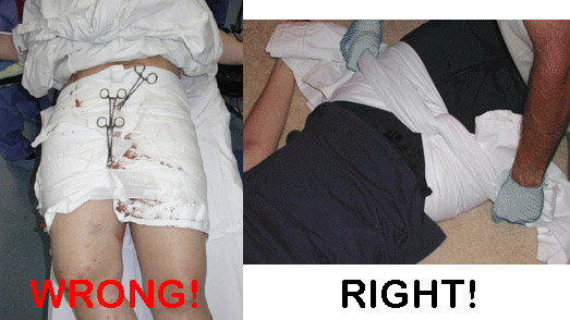
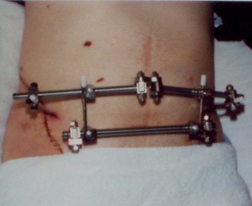
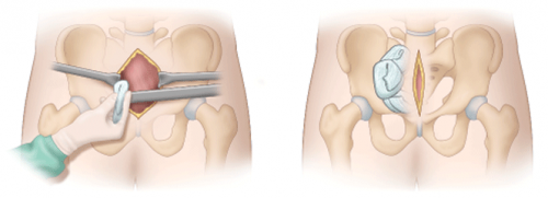
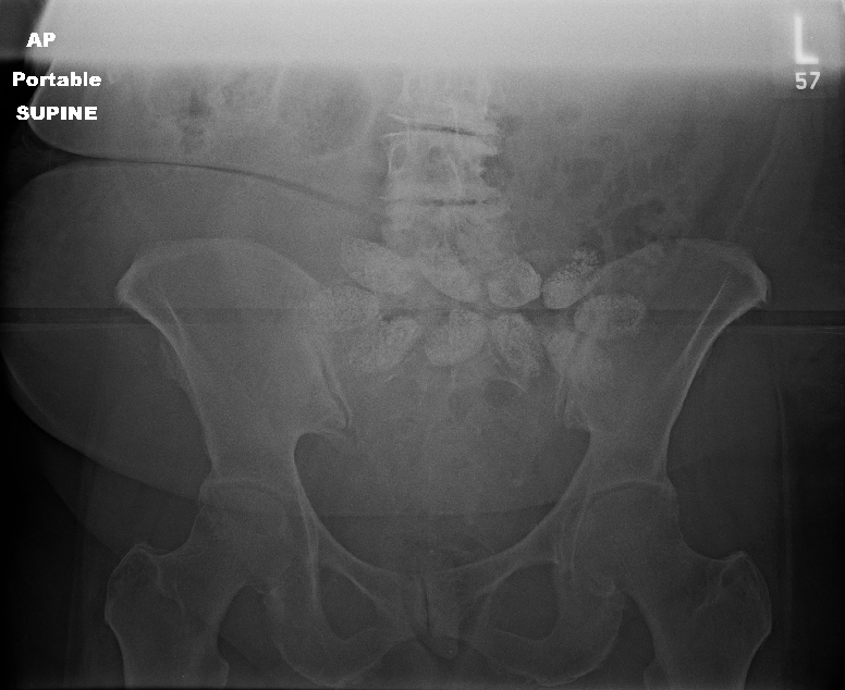
Angioembolization requires the presence of a special interventional radiology team and a reasonably stable patient. The procedure can take some time, and the IR suite is not really the place to house an unstable patient. Preperitoneal packing requires a reasonably stable pelvis to hold the packs in place for optimal tamponade, which may require application of an external fixator at the time of the procedure.

But is one better than the other? A number of relatively small studies have been performed, which means that it is time to synthesize them and see if some clearer answers can be found. The trauma group in Newcastle, Australia did just this. They performed a systematic search of the literature, analyzing the impact of each procedure on in-hospital mortality.

Here are the factoids:

  • A total of 18 studies met the authors’ inclusion criteria: 6 studies on AE, 9 studies of PPP, and 3 that compared them to each other
  • ISS was significantly higher in the PPP group vs AE (41 vs 36)
  • Average time to OR in the PPP patients was 60 minutes vs 131 minutes to IR in the AE group (statistically significant)
  • A quarter (27%) of the PPP patients did not get adequate hemorrhage control and required AE
  • In-hospital mortality in the PPP papers was 23% vs 32% in the AE research
  • Mortality in the papers that compared AE directly to PPP wasno different

Bottom line: What does this all mean? Is packing “better” than embolization? The simple answer is that we don’t know yet. Due to the way this study was performed, it is not possible to tease out all of the possible confounders. 

We are taught that control of hemorrhage is paramount. The time to definitive management in the AE group was twice that of the PPP patients. This could have a major impact on mortality. Two hours of bleeding can certainly kill. And the lower mortality in the PPP group occurred even though their injury severity was higher.

Many trauma centers have both of these interventions available to choose from. How should we approach their use? Unfortunately the literature is still to scarce to come to strong conclusions. Until we have better research to learn from, I suggest the following:

  • Time is of the essence. Which procedure can you get the fastest? In many cases, this will be preperitoneal packing since it’s just a trip to your trauma OR, which should be ready and waiting. If you have an IR team standing by or available very quickly, you could consider them first.
  • Pay attention to hemodynamic stability. An IR suite is no place for an unstable patient. The resuscitation equipment is not on par with the OR, and one never knows exactly how long the procedure will last.
  • If you have a hybrid room, use it! This is the ideal situation. The surgeon can start the PPP while the orthopedic surgeon applies a fixator. And the radiologist can be preparing to finish it off with a quick squirt as soon as they move away from the groin.
  • The use of one does not rule out the other. If one fails and the patient has increasing fluid and blood requirements move immediately to the other procedure to try to get control.

Reference: Preperitoneal packing versus Angioembolization for the initial management of hemodynamically unstable pelvic fracture – A Systematic Review and Meta-Analysis. J Trauma, publish ahead of print, Jan 4 2022, doi: 10.1097/TA.0000000000003528.


Options For Hemorrhage Control From Pelvic Fracture

We’ve come a long way in our available treatments to slow or stop bleeding from pelvic fractures. Let’s work our way through the list in today’s post, then look critically at two of the newcomers in the next one.

Pelvic binders. Long ago, these were just sheets wrapped around the patient and secured with clamps.

They were rather crude, as you can see. So of course, several enterprising companies began to offer commercial binders that were easier to place and secure.

Of note in the photo above, the wrap on the left is totally wrong. It is too wide and extends too high, so will not provide effective compression. The image on the right shows proper placement low across the greater trochanters. It is also not secured using metal clamps which may interfere with x-ray imaging.

External fixation of the pelvis. This usually involved a call to your friendly orthopedic surgeon. It could be applied in either the trauma bay or the operating room.

This image also shows improper technique. The horizontal bar should be angulated downwards over the pubis so it will not interfere with the trauma surgeon’s approach to laparotomy.

Internal pelvic packing + internal iliac artery ligation. Since surgeons didn’t have many other good tools, they could actually operate! Unfortunately, neither of these worked terribly well. The laparotomy pads could decompress upwards out of the pelvis and the internal iliac arteries have lots of collateral branches that permit ongoing bleeding from pelvic bones.

Angioembolization. Arterial bleeding from the pelvis occurs more often than you think (upwards of 50% of major pelvic injuries). Angiography and embolization can work very well. Unfortunately they are not suitable for unstable patients since IR suites are poor resuscitation areas. Many trauma centers do not have hybrid operating rooms where hemodynamically compromised patients can be taken for combined IR and open procedures if needed. So unstable patients must go to a regular OR first to attempt stabilization.

Preperitoneal packing. This is the new OR procedure kid on the block. Instead of placing packs in the pelvis, they are placed next to the broken pelvic bones but just outside the peritoneum. This permits better tamponade, and the intraperitoneal viscera push out against the packs to help decrease bleeding.

Zone 3 REBOA. And this is the very newest kid on the block. The balloon tipped catheter is inserted to a level above the aortic bifurcation but below the visceral and renal vessels. This is essentially a non-selective, temporary ligation of not just the internal iliac arteries, but everything distal to the aorta. It can be performed in the ED to dramatically slow blood loss, providing more time to get the patient to the OR where more definitive hemorrhage control can be provided (using many of the above techniques).

In my next post, I’ll take a closer look at the effectiveness of preperitoneal packing vs angioembolization.

Best Practice: Use of CT Scan In Trauma Activations – Part 2

In my last post, I described how the unscheduled and random use of CT scan in trauma activations can interfere with normal radiology department workflow, creating access problems for other emergency and elective patients. Today, I’ll detail a project implemented at my hospital to analyze the magnitude of this problem and try to resolve it.

We started with a detailed analysis of how the scanner was being used for trauma activation patients. Regions Hospital has a single-tier trauma activation system, with no mechanism of injury criteria other than penetrating injury to the head, neck, and torso. There were usually about 850highest-level activations per year at the time, and traditionally the CT scanner had been “locked down” when the activation is announced. The CT techs would complete the current study on the table, then hold the scanner open until called or released by the trauma team.

Since we are a predominantly blunt trauma institution, we scan most stable patients. Our average time in the trauma bay is a bit less than 20 minutes. Add this time to the trauma activation prenotification time of up to 10 minutes, and the scanner has the potential to sit idle for up to half an hour. And in some cases when scan is not needed (minor injuries, rapid transport to OR) the techs were not notified and were not aware they could continue scanning their scheduled cases.

A multidisciplinary group was created and started with direct observation of the trauma activation process and a review of chart documentation and radiology logs. On average it was calculated that the scanner was held idle for an average of 17.9 minutes too long. This is more than enough time to complete one, or even two studies!

A new process was implemented that required the trauma team leader to call out to the ED clerk placing orders for the resuscitation 5 minutes before the patient would be ready for scan. I still remember the first time this happened to me. I was so used to just packing up and heading to scan, I got a little irritated when told that I hadn’t made the 5-minute call. But it’s a good feedback loop, and I never forgot again!

We studied our workflow and results over a 9-week period. And here are the factoids:

  • The average CT idle time for trauma activations before the project was 17.9 minutes
  • This decreased to an average idle time of 6.4 minutes during the pilot project
  • Total idle time for all activations was 8.3 hours, but would have been 36 hours under the old system
  • A total of 28.6 hours were freed up, which allowed an additional 114 patients to be scanned while waiting for the trauma activation patients

This was deemed a success, and the 5-minute rule is now part of the routine flow of our trauma activations. We rarely ever have to wait for CT, and if we do it’s usually due to the team leader not thinking ahead.

Bottom line: This illustrates the processes that should be used when a quality problem surfaces in your program:

  • Recognize that there is a problem
  • Convene a small group of experts to consider the nuances
  • Generate objective data that describes the problem in detail
  • Put on your thinking caps to come up with creative solutions
  • Test the solutions until you find one that shows the desired improvement
  • Be prepared to modify your new systems over time to ensure they continue to meet your needs

Best Practice: Use of CT Scan In Trauma Activations – Part 1

Computed tomography is an essential part of the diagnostic workup for many trauma patients. However, it’s a limited resource in most hospitals. Only so many scanners are affordable and available.  Typically, trauma centers have a scanner located in or very near the trauma bay, which makes physical access easy. Others may be located farther away, which can pose logistical and safety issues for critically injured patients.

Even if the CT is close to the ED, availability can be an issue. This availability applies not only to trauma scans, but to others as well. There is an expectation that CT be immediately available when needed for trauma activation patients. However, chances are that the same scanner is also used for high priority scans for services other than trauma, such as stroke evaluation.

Who gets the scanner first? Obviously, many trauma patients need rapid diagnosis for treatment of their serious injuries. But a fresh stroke patient also has a neurologic recovery countdown clock running if they might be eligible for lytic administration.

And don’t forget that trauma and stroke aren’t the only services vying for that scanner. The hospital undoubtedly has a stream of elective scans queued up for other in-house patients. Every urgent or emergent scan needed for trauma sets the elective schedule back another 30 minutes or more.

How does your trauma center manage CT scan usage for trauma? The vast majority essentially lock it down at some fixed point. This is typically either upon trauma activation, or at patient arrival. The former is very common, but also very wasteful because there can be a significant wait for the patient to actually arrive. Then add on the time it takes to complete the trauma bay evaluation. Up to an hour may pass, with no throughput in the CT scanner. This can be a major work flow headache for your radiology department.

Is there another way? My center was one of those that stopped the scanner after the current patient was finished at the time the trauma activation was called. We have two scanners just 30 feet from the trauma bays, so one could continue working while the other was held. However, this cut their throughput by 50% for roughly half an hour. We recognized that this was a creating a problem for the whole hospital, so we worked with the radiology department to come up with a better way.

In my next post I’ll detail the new system we implemented, and provide data showing the real impact of this new system on CT scan productivity.

What The Heck?! The Answer!

In my last post, I described an elderly pedestrian struck by a car. During the trauma activation, routine chest and pelvic x-rays were obtained. Here was the pelvic image:

Note the odd oval densities across the center of the x-ray. What the heck? What are they?

There are two choices: they are either inside or outside the patient. We had already removed her clothes, so it wasn’t something she was carrying. And if it was inside, we would be able to identify it on the CT scan we had ordered.

But in this case, the x-ray was done early in the secondary survey. Specifically, we did it before we rolled our patient and examined her back.  When we did, here is what we found:

Only it wasn’t in the box. Or on her neck. This one was stuck on her lower back, but not in her clothes. She was suffering from lower back pain, and applied one of these on a daily basis for comfort. We had not rolled the patient prior to the pelvic x-ray.

The pods on these thermal wraps contain a mixture of iron, sodium chloride, sodium thiosulfate, water, charcoal, and sodium polyacrylate that heat up when removed from their package and exposed to oxygen. The iron renders it somewhat radio-opaque, hence their appearance on the x-ray. We did peel it off prior to CT since it would probably create a significant amount of scatter which would degrade the image.

Should we have waited a few more minutes to get the image until we had rolled and examined the back? This is a judgment call. Since our trauma team moves quickly, we are typically ready to head to the scanner in 15 minutes. In order to improve overall CT scan throughput, we have adopted a 5-minute advance notice policy.  To accomplish this, we don’t want to wait until the very end of the resuscitation to get x-rays. That would end up slowing down our process.

You may feel differently about the timing of the images, or you may have a different method of sequencing your CT scanner. Whatever works best for you. But remember, all trauma patients need to be completely undressed and all of their surfaces, nooks, and crannies inspected before they leave the emergency department!