All posts by The Trauma Pro

Nail In The Neck: The Operation

We’ve made sure that our victim of the nail gun to the neck did not need an emergent operation. Vitals are stable, there’s no uncontrolled hemorrhage, and the patient is neurologically intact. We’ve imaged him using CT angiography, and the nail does not appear to have injured any vital structures.

How do we get it out of there? There are two things that need to be considered: where and how.

Rule of thumb: If a foreign body is located anywhere near vital structures, take it out in the OR, no matter how good you think the imaging is. It may be tempting to just pull it out in the ED, but resist! CT scans look so good, and they are so detailed, but they are not perfect. The ED does not have the equipment, personnel, or lighting necessary if something goes awry.

Rule of thumb: Use all information available to plan the removal procedure. In this case, the head of the nail is to the patient’s right. Therefore, it must be removed from the right side. The CT shows that the nail passes very close to the posterior pharynx, so it will need to be evaluated during the procedure.

This patient was taken to the operating room. During the intubation, direct laryngoscopy was carried out to carefully inspect the entire pharynx and larynx. No evidence of penetration was seen. The entire neck, face, and upper chest were prepped and draped (I like to go overboard in trauma cases; you never know what is going to happen). Fluoroscopy was available.

The classic operation would have been to make an incision along the sternocleidomastoid on the right side. The nail head would be approached directly. Since long, thin objects can be notoriously difficult to locate, fluoro can be very helpful. The exact position with respect to the carotid and jugular can be ascertained. Then the nail head is grasped with a clamp, and the nail gently pulled out along its axis. A nice, long wait for any evidence of bleeding should occur. The area can then be irrigated and the incision closed. Skin antibiotics can be given postop, but only one dose at most.

Having said that, I opted for a different approach based on an old blog post here. Tune in to the final post in this series to see what I really did.

Nail In The Neck: Part 2

This case involves an accidental nail gun injury to the neck. The patient is hemodynamically stable, neurologically intact, the airway is patent and not threatened, and there is no apparent hematoma. There is a small puncture near the sternocleidomastoid muscle on the right, fairly high on the neck. The nail is not palpable on either side. And the patient only complains of a little discomfort when he swallows.

What to do? First, the patient has passed all the initial decision points that would send us straight to the OR (ABC problems in ATLS jargon). But, per physical exam and initial imaging, the nail must obviously come out. We just have to figure out what we need to know before we take it out, and determine the best way to retrieve it.

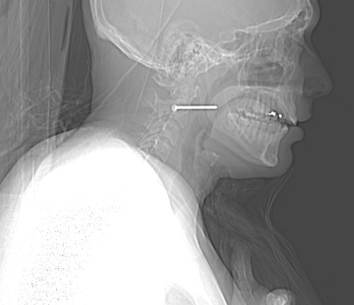
Given the patient’s stability, additional imaging will be helpful. Views in different planes, and details of what the nail might have passed through will be invaluable. The recommended study is a CT angio of the neck. This will give good information about nearby structures and the vasculature. And software reconstructions will provide good 2D/3D information for removal planning. Here’s a lateral view.

image

The nail is located in front of the body of C2. It appears to be high enough to be near the pharynx, but well above trachea and esophagus. You can also see that the nail entered a little posteriorly, and travels right to left and forward.

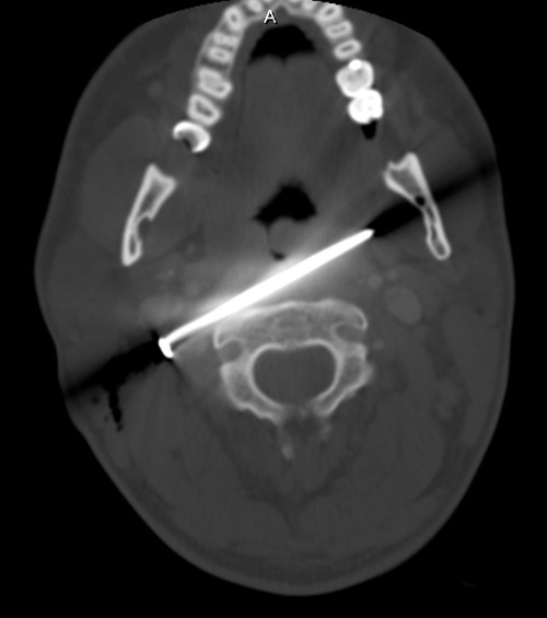
Here’s a representative CT slice.

image

The nail enters behind the carotids (just above the bifurcation) and IJ on the right, and ends anterior to them on the left. It passes very close to the posterior pharynx. So neurovascular structures are intact, and the aerodigestive tract is a maybe (back of the pharynx).

Obviously, this thing has to come out. The question is, how to do it? For you surgeons out there, tell me your choice of approach, incision, and instrumentation. Tweet/X or leave comments! Answers in the next post.

What Would You Do? Nail In The Neck

Here’s a very interesting case for you. A construction worker was carrying an object inside a building WHILE HOLDING HIS NAIL GUN! As he passed through the door, his elbow hit the frame and he brushed his neck with the business end of the gun. Guess what happened?

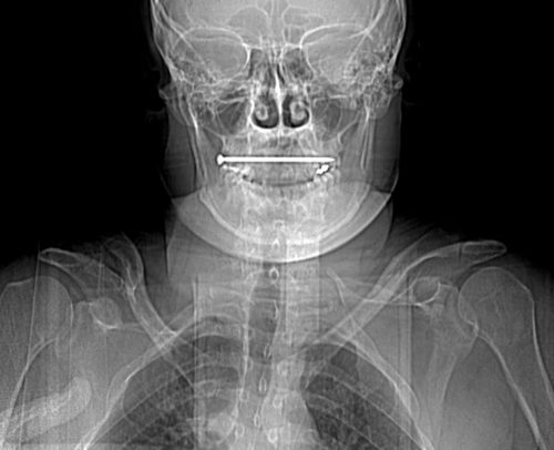
He experienced sharp pain, then noted pain every time he swallowed. He checked himself out in the mirror, and there was a small puncture wound in the right side of his neck. He presented to his local ED, with the complaints just noted. He was hemodynamically stable and neurologically intact. His airway was patent, and he had minimal pain. The following image was obtained. The nail measures about 6cm in length.

Let me know (by X or comments) what additional information you think you need. I’ll discuss that in my next post. Then we’ll figure out what to do about this problem.

Hypotensive Patient? You’ve Got 90 Seconds!

You’re running a trauma activation, and everything is going great! Primary survey – passed. Resuscitation – lines in, fluid going. You are well into the exam in the secondary survey.

Then it happens. The automated blood pressure cuff shows a pressure of 72/44. But the patient looks so good!

You recycle the cuff. A minute passes and another low pressure is noted, 80/52. You move the cuff to the other arm. Xray comes in to take some pictures. You roll the patient. 76/50. Well, you say, they were lying on the cuff. Recycle it again.

A minute later, the pressure is 56/40, and the patient looks gray and is very confused and diaphoretic. It’s real! But how long as it been real? An easy 5 minutes have passed since the first bad reading.

Bottom line: Sometimes it’s just hard to believe that your patient is hypotensive. They look so good! But don’t be fooled. If you get a single hypotensive reading, STOP! You have 90 seconds to figure out if it’s real, so don’t do anything else but. Check the pulse rate and character with your fingers. Do a MANUAL blood pressure check. It’s fast and accurate. If you have the slightest doubt, ASSUME IT’S REAL.

Don’t get suckered into trying to figure out what’s wrong with the cuff despite how good your patient looks. Remember, your patient is bleeding to death until proven otherwise. And it’s your job to prove it. Fast!

The Lowly Blood Pressure Cuff: Is It Accurate?

Yesterday, I described how the typical automated oscillometric blood pressure cuff works. We rely on this workhorse piece of equipment for nearly all pressure determinations outside of the intensive care unit. So the obvious question is, “is it accurate?”

Interestingly, there are not very many good papers that have ever looked at this! However, this simple question was addressed by a group at Harvard back in 2013. This study utilized an extensive ICU database from 7 ICUs at the Beth Israel Deaconess Medical Center. Seven years of data were analyzed, including minute by minute blood pressure readings in patients with both automated cuffs and indwelling arterial lines. Arterial line pressures were considered to be the “gold standard.”

Here are the factoids:

  • Over 27,000 pairs of simultaneously recorded cuff and arterial line measurements from 852 patients were analyzed
  • The cuff underestimated art line SBP for pressures at or above 95 torr
  • The cuff overestimated SBO for pressures below 95 torr (!)
  • Patients in profound shock (SBP < 60) had a cuff reading 10 torr higher
  • Mean arterial pressure was reasonably accurate in hypotensive patients

sbp-cuff-v-aline

Bottom line: The good, old-fashioned automated blood pressure cuff is fine for patients with normal pressures or better. In fact, it tends to understimate the SBP the higher it is, which is fine. However, it overestimates the SBP in hypotensive patients. This can be dangerous! 

You may look at that SBP of 90 and say to yourself, “that’s not too bad.” But really it might be 80. Would that change your mind? Don’t get suckered into thinking that this mainstay of medical care is perfect! And consider peeking at the mean arterial pressure from time to time. That may give you a more accurate picture of where the patient really is from a pressure standpoint.

Reference: Methods of blood pressure measurement in the ICU. Crit Care Med Journal, 41(1): 34-40, 2013.