All posts by The Trauma Pro

How To Remove A Tourniquet

Tourniquets had been banished for several decades due to the misconception that they caused more harm than good. But thanks to the experience of the US military, they have made a resurgence again in civilian use. If handled properly, they can literally be a life-saver.

More and more often, our prehospital trauma professionals are applying a tourniquet in the field. The question once they arrive in your trauma bay is “now what?”

Well, obviously it’s got to come off. But there is a lot of nuance around how to do that. And I don’t just mean the technical aspects of releasing it. It’s important to understand what injuries your patient has, and what the capabilities of your trauma center are first. Here is a framework to help you think through the details.

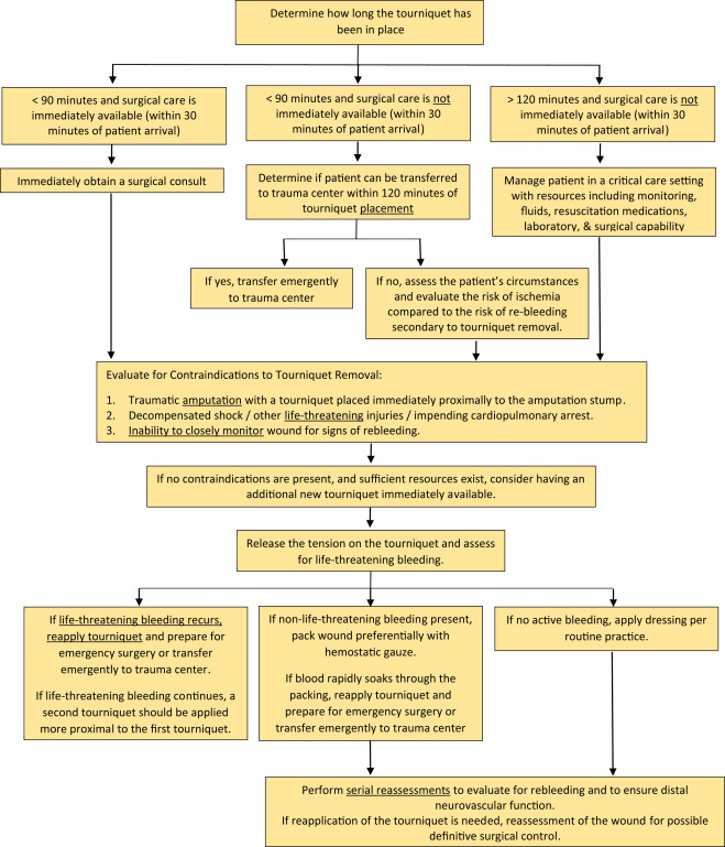
  1. How long has the tourniquet been up? Hopefully that has been recorded somewhere, or written on the tourniquet. If you don’t know exactly, assume that medics applied it upon arrival at the scene.
    1. If < 90 minutes and you have surgical support available, call the surgeon! If they believe the patient needs to be in the OR right away, make it happen.
    2. If < 90 minutes and you do not have surgical support, transfer your patient ASAP to a center that has it. If the transfer will take more than 2 hours (due to distance / weather and not a slow transfer on your part, consider dropping the tourniquet as described below.
    3. If > 120 minutes regardless of transfer status, consider dropping the tourniquet as described below.
  2. Is there a contraindication to removal?
    1. Traumatic amputation with the tourniquet nicely placed just proximal to an amputation stump. It may slip off after releasing the tension.
    2. Decompensated shock or near arrest. The patient is trying to die and the tourniquet is helping to prevent them from doing just that.
    3. Inability to closely monitor for rebleeding. If the patient needs to be transported in a relatively unsupervised setting, new bleeding may not be treatable.

If there are no contraindications and there is a need to at least temporarily release the tourniquet, then prepare your area appropriately.  Ideally, this should be done in an OR or ICU, but that is not always practical. Otherwise, make your trauma bay look like one. Make sure you have at least one new tourniquet in case the old one can’t be reapplied for some reason. Ensure there is plenty of hemostatic gauze and dressing materials. Have the crash cart nearby and make the ACLS drugs readily available, just in case.

Then release the tension on the tourniquet and note the time. Three things can happen:

  1. There is no bleeding. This happens about 80% of the time in my experience. Either there was no surgical bleeding in the first place, or it has clotted. Place a nice dressing that can be monitored easily.
  2. There is only “non-surgical” bleeding. This is typically oozing or pesky venous bleeding. These should be controlled with sutures or hemostatic dressings. Pressure dressings are also wonderful in the situation. Craft them carefully.
  3. Life threatening bleeding resumes. Reapply the tourniquet and get the patient to definitive care ASAP (OR or another center that has one).

Bottom line: There is very little magic to dealing with tourniquets on the receiving end. But get a very clear picture of what your patient needs and what your center has to offer them. If these factors don’t match up, initiate the transfer as fast as you possibly can. Otherwise move to your OR to fix the problem!

Reference: Removal of the Prehospital Tourniquet in the Emergency Department. J Emerg Med 60(1):98-102, 2020.

What GCS Should Trigger Trauma Activation?

For the most part, trauma centers are free to pick and choose their own trauma team activation trigger criteria. Typically, these are a mix of physiologic, anatomic, and mechanistic items. However, the American College of Surgeons Committee on Trauma mandates that either seven (Orange Book) or eight (Gray Book) specific criteria must present in every center’s highest-level activation list.

One of these mandatory criteria is a Glasgow Coma Scale (GCS) score of eight or less. The reason is that this level denotes a severe brain injury and as patients approach it they are less and less able to protect their own airway. Although this specific GCS is a minimum, centers are free to choose their own specific threshold as long as it is not any lower.

How does a center choose the “right” GCS? It seems straightforward, right? A mild TBI is defined as GCS from 13-15. These patients have only lost one or two points in their eye-opening, verbal, and motor scores and are relatively unlikely to have a significant lesion in their head or an airway issue.

At the other end of the spectrum is the severe TBI, with a GCS of 3-8. These are a chip shot, with the potential for severe injury and a frequently threatened airway. They demand rapid assessment and intervention, hence the required trauma activation.

But what about those patients with moderate TBI with a GCS from 9-12? They obviously have a higher risk for serious intracranial injury. And as the GCS declines, the patient’s ability to protect their airway decreases. At some point between those GCS scores, most clinicians hit their own internal trigger to provide a definitive airway.

So what do actual trauma centers choose as their threshold? I conducted an informal survey of my readers, asking them to provide their specific GCS threshold.

Here are the factoids:

  • A total of 147 trauma centers of all levels responded
  • They were located in the United States, Germany, Saudi Arabia, and Singapore
  • This chart shows the number of centers that selected a threshold less than or equal to the GCS on the horizontal axis:


  • Nearly a third of centers (30%) adhere strictly to the ACS criterion of 8
  • Another 22% use a threshold of 9, possibly to avoid any confusion from having a “less than or equal to” criterion
  • There is another bump on the curve at 13, with 20% using this threshold

Bottom line: A little more than half of centers use a GCS threshold of 8 or 9 as their TTA trigger. This meets the ACS criteria, but could potentially leave a few airways unprotected from time to time. Only about 5% of centers use the higher GCS levels with the exception of GCS 13. That seems to be another popular one.

Which one is right for you? GCS 8 will always work because it is the minimum requirement. My own personal threshold trends higher. I would rather be called to an activation and apply my own judgement rather than come running only when the patient needs to be intubated followed by a trip to the OR for craniotomy.

You will need to work with your emergency physicians, trauma surgeons, and neurosurgeons to determine their collective comfort levels. It comes down to a balance between safety and unnecessary intubation. Look at your own center’s experience and pick a threshold that achieves a proper balance of overall patient safety.

Maxillofacial CT Scans In Children

Facial trauma is common, especially in children. And the use of CT scan is even more common, unfortunately for children. What happens when these two events meet?

I’ve noted that many trauma professionals almost reflexively order a face CT when they see any evidence of facial trauma. This ranges from obvious deformity to lacerations to mere contusions. This seems like overkill to me, since most of the face (excluding the mandible) is visualized with the head CT that nearly always accompanies it.

Finally, someone has actually examined the usefulness of the facial CT scan! The trauma group at Albany collaborated with four other Level I trauma centers, performing a retrospective chart and database review of children (defined as less than 18 years old) who underwent both head and maxillofacial CT scans over a five year period. They excluded penetrating injuries and bites. The concordance of facial fractures seen on head CT vs face CT was evaluated.

Here are the factoids:

  • A total of 322 patients with facial fractures was identified, and the most common mechanisms were MVC, pedestrian struck, and bicycle crash
  • Fractures on head CT matched the facial CT in 89% of cases
  • Of the 35 discordant cases, 21 of the head CTs missed nasal fractures, 9 mandibular fractures, 3 orbital fractures, and 2 maxillary fractures
  • Of those 35 cases, only 7 required operative intervention: 6 mandible fractures and 1 maxillary fracture

The authors concluded that the use of head CT alone with a good clinical exam detects nearly all facial fractures requiring repair.

Bottom line: Although this study confirms my own personal bias and experience, it suffers from the usual problems associated with retrospective studies and small numbers. Nonetheless, the results are compelling. This study provides a way to identify nearly all significant fractures while minimizing radiation to the ocular lens, thyroid, and bone marrow.

The key is a good physical exam, as usual. Inspection of the teeth, occlusion testing, and manipulation of the mandible and maxilla should identify nearly all fractures that might require operation.

Once the exam is complete, a standard head CT should be obtained. Identification of displaced fractures on the head CT should prompt a consult to your friendly facial surgeon to see if they really need additional imaging to determine if the fracture requires operation. Frequently, the head CT images are sufficient and nothing further is required.

Here is the algorithm the authors recommend. Although designed for children, it should work for adults just as well.

Reference: Clinical and radiographic predictors of the need for facial CT in pediatric blunt trauma: a multi-institutional study. Trauma Surg Acute Care Open 2022;7:e000899.

The Ultimate Distracting Injury?

By now, we are all very familiar with the concept of the distracting injury. Some of our patients sustain injuries that are so painful that they mask the presence of others. The patient is so distracted by the big one that others just slip their notice.

This concept has been notoriously difficult to test, but there is a reasonable amount of data that suggests it is true. One of the more common and disturbing injury patterns occurs when there is a significant amount of chest wall trauma. When there are fractures focused around the upper chest, cervical spine injuries may be masked, then missed during the exam by trauma professionals.

I’d like to introduce a new concept: the ultimate distracting injury. This goes beyond an injury distracting the patient from another painful problem.

The ultimate distracting injury is one that is so gruesome that it distracts the entire trauma team! It could actually be so distracting that the team might miss multiple injuries!

It’s just human nature. We are drawn to extremes, and that goes for trauma care as well. And it doesn’t matter what your level of training or expertise, we are all susceptible to it. The problem is that we get so engrossed (!) in the disfiguring injury that we ignore the fact that the patient is turning blue. Or bleeding to death from a small puncture wound somewhere else. We forget to focus on the other life threatening things that may be going on.

What are some common ultimate distracting injuries?

  • Mangled extremity
  • Traumatic amputation
  • Impalement
  • Severe soft tissue injury

How do we avoid this common pitfall? It takes a little forethought and mental preparation. Here’s what to do:

  • If you know in advance that one of these injuries is present, prepare your crew or team. Tell them what to expect so they can guard against this phenomenon.
  • Quickly assess to see if it is life threatening. If it bleeds or sucks, it needs immediate attention. Take care of it immediately.
  • If it’s not life threatening, cover it up and focus on the usual priorities (a la ATLS, for example).
  • When it’s time to address the injury in the usual order of things, uncover, assess and treat.

Don’t get caught off guard! Just being aware of this common pitfall can save you and your patient!

When To Call Your Ophthalmology Consultant

And yes, another consultant reference card. As I wrote previously. we sometimes overuse our consultants and call then at inappropriate times. So what if we diagnose an injury in their area of expertise at 2 am? Does it need attention or an operation before morning? If not, why call at that ungodly hour?

Let’s use our consultants wisely! I’ve listed most of the common eye diagnoses that trauma professionals will encounter. There is also an indication of what you need to do, and exactly when to call your consultant.

Unfortunately, this one won’t fit on a 3×5 index card that you can keep in your pocket. I’ve included a printable pdf file, as well as the original Microsoft Word file in case you want to make a few modifications to suit your own hospital.

Enjoy!

When to call Ophthalmology reference card (pdf)

When to call Ophthalmology MS Wordfile (docx)