All posts by The Trauma Pro

The Impact Of NSAIDs On Fracture Healing

In my last post I discussed some of the basic effects of NSAIDs on bone healing. Now let’s see if theory applies to practice.

In 2003, several papers brought to light possible interactions between these drugs and fracture healing. Specifically, there were questions about these drugs interfering with the healing process and of increasing the number of delayed unions or nonunions. But once again, how convincing were these papers, really?

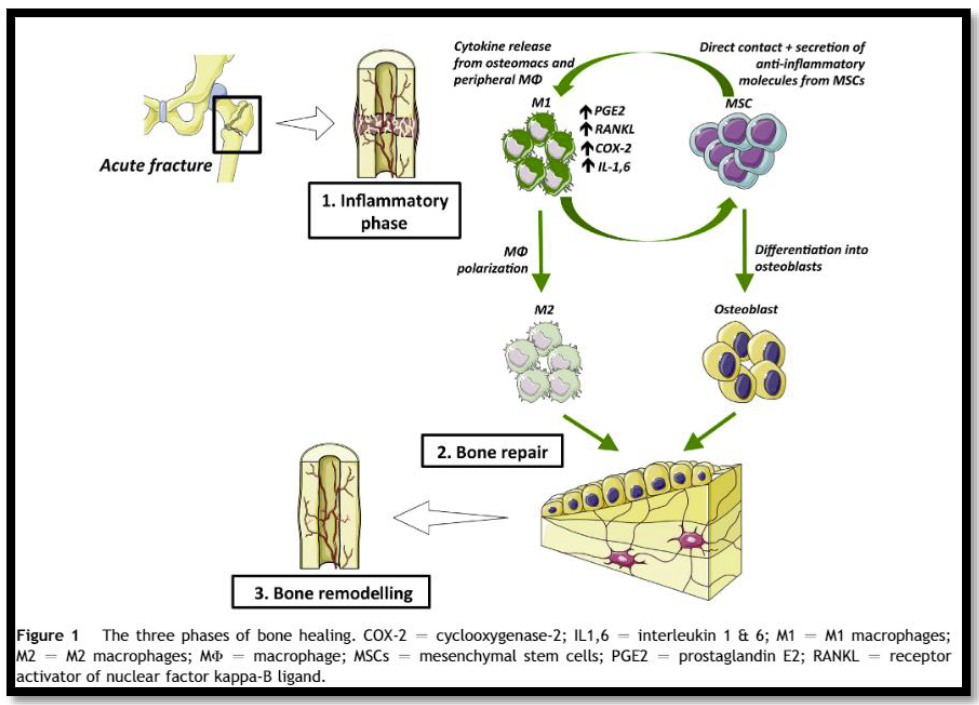
It would seem to make sense that NSAIDs could interfere with bone healing. The healing process relies heavily on the regulation of osteoblast and osteoclast function, which itself is regulated by prostaglandins. Since prostaglandins are synthesized by the COX enzymes, COX inhibitors like the NSAIDs should have the potential to impair this process. Indeed, animal studies in rats and rabbits seem to bear this out.

But as we have seen before, good animal studies don’t always translate well into human experience. Although a study from 2005 suggested that NSAID administration in older patients within 90 days of injury had a higher incidence of fracture nonunion, the study design was not a very good one. It was equally likely that patients who required these drugs in this age group may have been at higher risk for nonunion in the first place.

A meta-analysis of human studies was performed in 2011. Out of 558 potential studies, only 5 met criteria review. (This is yet another reminder of the sheer amount of sub-par research out there.) The authors found that short-term use (< 14 days) of normal dose NSAIDS was not associated with non-union. High doses of ketorolac (> 120mg/day) and diclofenac sodium (> 300mg total) did have an association. But remember, this does not show causation. There are many other factors that can impede healing (smoking, diabetes, etc).

A study from 2016 examined the effect of ketorolac administration on fracture healing in patients undergoing repairs of femoral and tibial fractures. It did not find an association between non-union and ketorolac, but did find one with smoking. Unfortunately, the study was small (85 patients given ketorolac, 243 controls without it). It probably does not have the statistical power to detect any difference with the NSAID. A power analysis was not provided in the methods section.

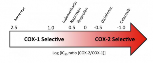
Bottom line: Once again, the animal data is clear and the human data less so. Although there are theoretical concerns about NSAID use and fracture healing, there is still not enough solid risk:benefit information to abandon short-term NSAID use in patients who really need them. NSAIDs can and should be prescribed in patients with short-term needs and simple fractures, and consider COX-1 specific drugs like ketorolac while your patient is in the hospital. And we do have some evidence that high-dose NSAIDs may have some impact, so stick to the usual doses for just as long as they are needed for pain management.

References:

  1. Effects of nonsteroidal anti-inflammatory drugs on bone formation and soft-tissue healing. J AM Acad Orthop Surg 12:139-43, 2004.
  2. Effect of COX-2 on fracture-healing in the rat femur. J Bone Joint Surg Am 86:116-123, 2004.
  3. Effects of perioperative anti-inflammatory and immunomodulating therapy on surgical wound healing. Pharmacotherapy 25:1566-1591, 2005.
  4. Pharmacological agents and impairment of fracture healing: what is the evidence? Injury 39:384-394, 2008.
  5. High dose nonsteroidal anti-inflammatory drugs compromise spinal fusion. Can J Anaesth 52:506-512, 2005.
  6. Nonsteroidal Anti-Inflammatory Drugs and Bone-Healing: A Systematic Review of Research Quality. JBJS Rev 4(3), 2016.
  7. High-dose ketorolac affects adult spinal fusion. Spine 36(7):E461-E468, 2011.
  8. Ketorolac administered in the recovery room for acute pain management does not affect healing rates of femoral and tibial fractures. J Orthop Trauma 30(9):479-482, 2016.

NSAIDs And Bone Healing: How Do They Impact It?

The arguments about whether NSAID administration has any effect on bone healing continues to be argued by our orthopedic and spine surgery colleagues. In the early days of research in this area (about 20 years ago) there were concerns in animal models that there might be a problem. Apparently lots of rats and bunnies were suffering from fractures in those days.

But physiologically, how could NSAIDS do this? Here’s a simplified diagram of how the bone healing process works.

First, an acute injury occurs and macrophages and other cells move into the area to start the inflammatory process. COX-2 receptors are highly expressed on these cells, resulting in an increase in prostaglandin E2 (PGE2) production.

PGE2 then promotes proliferation of stem cells that differentiate into osteoblasts, which in turn begin forming bone to repair the injury.  In theory, if PGE2 is reduced in the healing area there is the possibility that bone formation may be impaired, leading to non- or malunion or refracturing.

Administration of NSAIDs can block COX-1 and COX-2 receptors throughout the body. This serves to decrease prostaglandin production and hence reduces inflammation and pain. Doesn’t it follow that giving these drugs should be bad for bone formation in patients with fractures?

Not so fast! There are a number problems with this argument. First, not all NSAIDs are created alike. Here is a chart that shows where the primary focus of COX inhibition is with some common NSAIDs.

Note how the common over-the-counter drugs affect both COX-1 and COX-2, yet there are some that are more selective. So the choice of drug may be relevant.

And we can’t assume that an in vitro effect in a Petri dish of cells actually carries over into the in vivo world. Many researchers rely initially on animal models to study drug effects in vivo. Predictions based on studies of rats and bunnies frequently do not pan out in humans.

We are left with only a theory based on an understanding of the basic mechanism of bone healing. Tune in to my next post where I discuss the research that’s been done in this field and whether it actually translates into human bone healing or not.

Massive Transfusion: What Ratios Are People Using?

Back in the old days (which I remember fondly), we didn’t pay too much attention to the ratio of blood to plasma. We gave a bunch of bags of red cells, then at some point we remembered that we should give some plasma. And platelets? We were lucky to give any! And to top it all off, we gave LOTS of crystalloid. Turns out this was not exactly the best practice.

But things have changed. Some good research has shown us that a nice mix of blood component products is good and too much crystalloid is bad. But what exactly is the ideal mix of blood products? And what is everybody else doing?

What are all the other trauma centers doing? An interesting medley of anesthesia and pathology groups from the University of Chicago, a Dallas-based anesthesia group, and a blood center in my home base of St. Paul, conducted a survey of academic medical centers back in 2016. They wanted to find out how many actually had a MTP and to scrutinize the details.

They constructed a SurveyMonkey survey and sent it to hospitals with accredited pathology residencies across the US. There were 32 questions in the survey, which asked for a lot of detail. As you can probably personally attest, the longer and more complicated the survey, the less likely you are to respond. That certainly happened here. Of 107 surveys sent out, it took a lot of nagging (initial email plus two nags) to get a total of 56 back.

Here are the factoids:

  • Most were larger hospitals, with 74% having 500 or more beds
  • All had massive transfusion protocols
  • Trauma center level: Level I (77%), Level II (4%), Level III (4%), Level IV (2%), no level (14%)
  • Nearly all (98%) used a fixed ratio MTP; very few used any lab-directed (e.g. TEG/ROTEM) resuscitation
  • Target RBC:plasma ratio: 1:1 (70%), 1.5:1 (9%), 2:1 (9%), other (9%)
  • Only 58% had the same RBC:plasma ratio in each MTP cooler
  • More than 86% had thawed plasma available (remember, these were generally large academic centers)
  • Half stored uncrossmatched type O PRBCs outside the blood bank, usually in the ED; only 1 stored thawed plasma in the ED
  • A total of 41% had more than one MTP (trauma, OB, GI, etc.)
  • 84% had some type of formal review process once the MTP was complete
  • About 68% had modified their MTP since the original implementation. Some increased or decreased ratios, expanded MTP to non-trauma services, decreased the number of units in each pack, changed to group A plasma from AB, or switched from ratio to TEG/ROTEM or back.

Bottom line: This is an intriguing snapshot of MTP practices around the country that is about six years old. Also remember, this is a somewhat skewed dataset. The survey was directed toward hospitals with academic pathology programs, not trauma centers. However, there is enough overlap that the results are probably generalizable. 

Most centers are (were) using MTP packs containing six units of PRBCs, and were attempting to achieve a fixed 1:1 ratio. Half of hospitals had the same number of units in each cooler, half varied them by cooler number. Nearly half had multiple flavors of MTP for different specialties. Very few used TEG/ROTEM during the initial phased of MTP. Most modified their MTP over time.

Unfortunately, I’ve not seen a similar survey repeated recently. I’m certain that practices have changed over time as our understanding of balanced resuscitation continues to advance. 

Finally, I’ve written quite a lot on most of these issues. See the links to my “MTP Week” series below.

Reference: Massive Transfusion Protocols: A Survey of Academic
Medical Centers in the United States. Anesth & Analg 124(1):277-281, 2017.

MTP week series:

First, There Was REBOA, And Now… GROA?!

REBOA (Resuscitative Endovascular Balloon Occlusion of the Aorta) is a “newer” resuscitative technique that has actually been around since the Korean War. It was first used to treat two injured soldiers, and although they ultimately died, it spurred research into the technique and its applications.

Balloon occlusion techniques were then adopted by vascular surgeons and were found to be useful as an adjunct in managing ruptured abdominal aneurysms. A slow trickle of studies on human use in trauma began to surface. But when autopsy studies carried out during the Gulf War showed that uncontrolled torso hemorrhage was a major cause of death, research in the technique exploded.

First, there were a rash of pig studies evaluating the feasibility of using a percutaneously placed occlusion device in the early 2010s. This transitioned to human studies around 2014, and after that we were off to the races. Over 100 papers on REBOA are now published each year.

REBOA has been shown to have some advantage in patients with abdominal or pelvic sources of bleeding. The catheter is inserted in the groin and the balloon inflated in one of two zones, depending on the location of the hemorrhage (see diagram below). For abdominal bleeding, it is inserted just above the diaphragm in Zone I. For pelvic bleeding, it is inserted below the takeoff of the visceral arteries and above the aortic bifurcation, in Zone III.

In the US, REBOA catheters are only inserted upon arrival to the hospital. There are a few random reports of field placement where a physician is part of the prehospital team. By definition, this technique is generally not available in austere environments, only upon arrival in the emergency department.

Researchers at the University of Michigan began looking for an alternative technique that could be applied in the field by non-physicians. They noted the close anatomic relationship of the distal esophagus, proximal stomach, aorta, and thoracic vertebrae, and designed a device to compress the aorta against the spine in this area.

They developed a prototype device which they named GROA (gastro-esophageal resuscitative occlusion of the aorta). It consists of a gastro-esophageal tube with an ovoid balloon, an air pump with pressure measurement device, and an external compression device. Here is a picture of the device:

And here’s a diagram of what it looks like when inserted:

The tube is inserted and the balloon inflated. The external compression device is then placed around the patient, with the top plate located over the epigastrium and the bottom plate under the patient. It is designed to apply anterior pressure over the balloon, but to avoid circumferential constriction of the abdomen.

Bottom line: This device is an interesting development in the balloon occlusion space. As with early studies of REBOA, GROA is currently being investigated using a pig model. If it appears to be beneficial, it will still be several years before it makes the jump to human subjects. If effective, this concept would allow prehospital providers to apply some degree of hemorrhage control when it originates in the abdominal cavity.

There are currently exactly three papers on this new technique, and I have included the references below if you are interested in reading them. I’m sure there are many more to come and it may eventually be competing with REBOA for journal space.

There is one consideration to be aware of when reading these papers that is similar to much of the research on REBOA. Two of the authors have a financial interest in the company that licenses the GROA technology. And in the most recent study, another one of the authors is an advisory board member for one of the manufacturers of REBOA catheters. For these reasons, it is less likely that they will publish papers that are not favorable to the product. So read critically!

References:

  • Gastroesophageal resuscitative occlusion of the aorta: Physiologic tolerance in a swine model of hemorrhagic shock, Journal of Trauma and Acute Care Surgery: December 2020 – Volume 89 – Issue 6 – p 1114-1123 doi: 10.1097/TA.0000000000002867
  • Gastroesophageal resuscitative occlusion of the aorta prolongs survival in a lethal liver laceration model, Journal of Trauma and Acute Care Surgery: May 2022 – Volume 92 – Issue 5 – p 880-889 doi: 10.1097/TA.0000000000003444
  • Tandem use of Gastroesophageal Resuscitative Occlusion of the Aorta followed by REBOA in a Lethal Liver Laceration Model, Journal of Trauma and Acute Care Surgery: June 10, 2022 –  doi: 10.1097/TA.0000000000003719

MRI And External Fixators

MRI is an indispensable tool for evaluation of spine and soft tissue trauma. However, a great deal of effort was be made to ensure that any patient scheduled for this test is “MRI compatible.” The fear is that any retained metallic fragments may move or heat up once the magnets are activated.

But what about trauma patients with external fixators? That is one big hunk of metal that is inserted deep into your patient. There are three major concerns:

  • Is the material ferromagnetic? If so, it will move when the magnets are activated and may cause internal injury. These days, there are many fixator sets that are not ferromagnetic, avoiding this problem.
  • Can currents be induced in the material, causing heating? This is not much of a problem for small, isolated objects. However, external fixators are configured in such a way that loops are created. The fluctuating magnetic fields can induce currents that in turn will heat the surrounding tissue. And thinner materials (narrow pins) result in more current and more heating.
  • Will the metal degrade image quality?

The biggest challenge is that there is no standard ex-fix configuration. Our orthopaedic colleagues get to unleash their creativity trying to devise the appropriate architecture to hold bones together so they can heal properly. This makes it difficult to develop standardized guidelines regarding what can and can’t go into the scanner.

However, there is a growing body of literature showing that the heating effects are relatively small, and get smaller as the distance from the magnet increases. And non-ferromagnetic materials move very little, if at all, and do not interfere with the image. So as long as nonferromagnetic materials are used, the patients are probably safe as long as basic principles are adhered to:

  • Other diagnostic options should be exhausted prior to using MRI.
  • Informed consent must be obtained, explaining that the potential risks are not completely understood.
  • The fixator must be tested with a handheld magnet so that all ferromagnetic components can be identified and removed.
  • All traction bows must be removed.
  • Ice bags are placed at all skin-pin interfaces.
  • The external fixator must remain at least 7cm outside the bore at all times.

Bottom line: MRI of patients with external fixators can be safely accomplished. Consult your radiologists and physicists to develop a policy that is specific to the scanners used at your hospital. 

Related posts: