All posts by The Trauma Pro

… And The Ultimate Retained Foreign Bodies

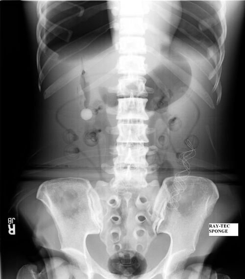
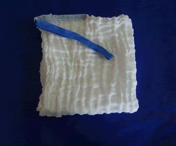
Sponges are unfortunately one of the most common retained foreign bodies. This is due to their small, flimsy nature. The surgeon usually looks at the obviously visible areas of the abdomen or other body cavity before closing. She can also feel around in the “nooks and crannies”, but sponges feel very similar to the other organs surrounding it.

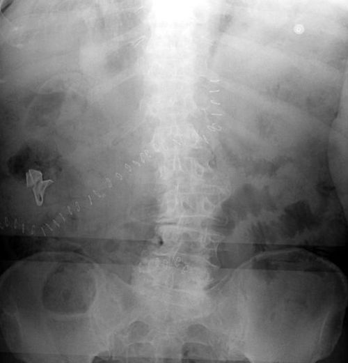
But what about more substantial items, like surgical instruments? Surely these are so obvious as to not leave behind?

Unfortunately, not so. Take a look at these items. This is a large pari of surgical forceps.

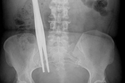
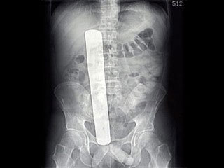
This is a malleable retractor, a long, thin sheet of pliable metal that can be bent to any desired shape.

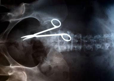
And finally, a pair of Metzenbaum scissor, a common surgical instrument for cutting tissue.

Bottom line: It doesn’t matter how small or large, anything can and will be left behind in emergent and trauma cases. Recognizing that this can occur, no matter how confident you are that it has not, is the key. Always count, but followup with an x-ray that covers all areas of the surgical field before closing.

Related post:

What Does A Retained Surgical Sponge Look Like?

It’s the bane of any surgeon’s existence. And the reason why OR personnel take such great pains to account for everything in the room. It is a catastrophe, and always a preventable one, when some piece of equipment goes missing and ends up left inside a patient.

A number of methods have been developed to try to eliminate this problem. They include careful counts, having someone record anytime anything is placed inside, x-rays, and most recently, RFID tags. 

After counting, x-ray is the most common way to try to find missing objects. One would think that these foreign bodies would be easy to see. Metallic instruments are rather easy to spot. But many trauma professionals, even those who work in the OR, have never seen what a positive image of a sponge actually looks like. So here they are. You should never miss one on an xray now.

Surgeons typically use two types of sponges in the OR: Ray-Tec sponges and standard lap pads. Ray-Tecs look like a 4×8 piece of gauze with a mysterious blue string woven throughout it. The string is the only part that shows up on x-ray, and it is very thin and somewhat hard to see. Here are some Ray-Tec sponges outside the body:

And here’s one that was left inside. Note the little squiggle in the left lower quadrant and how easy it is to over look.

On the other hand, a laparotomy pad is a 4×4 folded cloth pad that unfolds into a larger pad. It has a blue tag sewn in the corner, extending along one edge of the pad. Here’s what they look like:

And here’s one inside a patient. Note the irregular object in the right upper quadrant.

Bottom line: It’s important for anyone who works in the OR on any body part to be familiar with the appearance of these tags on x-rays. Since it’s generally impossible to get accurate counts before or after a trauma procedure, always image the involved body cavity looking for these telltale signs before closing the patient.

Note: These images taken from the internet. Patients not treated at Regions Hospital.

September Trauma MedEd Newsletter Released

The September newsletter is now available! Click the image below or the link at the bottom to download. This month’s topic is chest trauma.

In this issue you’ll find articles on:

  • Why I didn’t like finger thoracostomy
  • Advanced needle thoracostomy
  • Trochar vs needle for tension pneumothorax
  • Troubleshooting chest tube air leaks
  • Chest tube collection systems gone wild
  • Managing chest tube air leaks
  • Pneumothorax in children

Subscribers received the newsletter first last weekend. If you want to subscribe (and download back issues), click here.

Click here to download and/or subscribe.

Return To Baseline After Concussion

Here’s another interesting paper that was presented at the Congress of Neurological Surgeons. There’s a lot of attention being focused on the incidence and management of concussion during sporting events. An international Concussion in Sport Group has been meeting for over 10 years, contemplating classification and management of this injury. They are considering using age to modify management of concussion in young athletes.

The authors looked at their own experience with 200 adolescent and young athletes. They stratified by age (younger = 13-16 year olds, older = 18-22 year olds), with 100 in each group. They matched them by number of previous concussions, and all underwent baseline and post-concussion ImPACT testing. They specifically looked at the number of days needed to return to baseline.

Interestingly, they identified significant differences in recovery time. And strangely enough, the older players did better than the younger ones. Overall, 90% returned to baseline within a month. But the younger players took 2-3 days longer to recover than the older ones. 

Bottom line: Looks like the Concussion in Sport Group is right on! Usually in trauma, older folks do worse than younger ones, so we tend to treat them more carefully. Not so in youngsters with concussions. Sports medicine physicians need to realize that the younger brain takes longer to recover, and they should err on the safe side and keep them out of the game longer. Objective testing to help predict return to play is extremely helpful.

Related post:

Reference: Sport-Related Concussion and Age: Number of Days to Neurocognitive Baseline. Oral presentation 145 – Congress of Neurological Surgeons 2012.

Nursing: When Is Drain Output Too Bloody?

Trauma surgeons frequently place some type of drain in their patients, whether it be a chest tube, a damage control system, or a bulb suction drain near the pancreas. On occasion, nursing may become concerned with the character of the output, wondering if the patient is bleeding significantly. How can you tell if the output is too bloody?

First, most drains are in place to drain serous fluid which may have a little blood in it. Drainage that is mostly bloody is very uncommon from these drains, which are typically placed after orthopedic, spine or abdominal surgery. However, some drains are placed in areas where unexpected bleeding may occur, such as:

  • Damage control drain systems – as patients warm up, arterial sources that were not surgically controlled may open up
  • Pericardial drains – more common in cardiac surgery, not trauma
  • Chest tubes in patients with penetrating trauma

What should you do if you have concerns about your patient’s drain output?

  • Familiarize yourself with what kind of drain it is and what it should be draining
  • Look at the volume of output – it takes 500cc of pure blood to drop the patient’s hemoglobin by about 1 gram. Low outputs are not dangerous, even if it is pure blood.
  • Look at the change in output– if it is increasing significantly or changes color, call the physician to evaluate.
  • Look at the color of the output – most drainage ranges from clear to something like cranberry juice and appears to be partially transparent. Look carefully if it appears to be darker or more opaque, and compare it to the blood that you would see in a blood collection tube. Even the darkest drain output usually looks a little watery compared to whole blood. Bright red output needs to be evaluated by a physician.
  • If in doubt, check the fluid’s hematocrit. Whole blood has a hematocrit of 30% or more. Most bloody-looking drain output maxes out at about 5%. If the value is closer to whole blood, have a physician evaluate the patient.