All posts by The Trauma Pro

Single Incision Fasciotomy?

The concept of performing 4-compartment leg fasciotomy using only one incision is not a new one. Techniques using a lateral approach, either with or without fibulectomy, have been described as early as 1967.

A new paper describes a single-incision technique for compartment syndrome using a medial approach. The authors believe that going through the anterior compartment to release the deep posterior is quicker, uses a smaller flap, and avoids injury to the peroneal nerve. They reviewed their own experience over a 5 year period.

Here are the factoids:

  • 180 fasciotomies were performed for compartment syndrome, of which 30 were single-incision
  • 27 had associated fractures, 2 were due to soft tissue injury and 1 was spontaneous
  • There was a single wound infection, nerve injury, and patient with persistent pain. There were several tethered tendons and scars.
  • The number and types of complications were similar to traditional fasciotomy

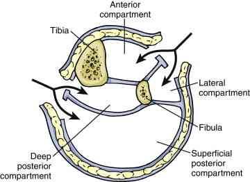
Traditional 4 compartment fasciotomy with 2 incisions. Source: gog.net.nz

Bottom line: Sounds great, right? Yes, it’s a small study, and statistically there is not enough power to show that it’s “better.” So if it’s not worse, and there is just one smaller incision, what’s wrong with it?

For me, the problem is that there is too much opportunity to perform an incomplete fasciotomy. The learning curve for single incision fasciotomy, either this one or the more traditional lateral approach, is steep. Seriously impaired patients who need fasciotomy are frequently not going to be awake any time soon, leaving the surgeon with no neurologic or pain exam.

My recommendation: read papers like this and smile. Then do the classic 2 incision operation, making sure that all compartments are completely released.

Related post:

Reference: A single incision fasciotomy for compartment syndrome of the lower leg. J Ortho Trauma, Publish ahead of print, January 21, 2016.

February Newsletter Released This Weekend – REBOA!

You’ve heard about it. You’ve read about. And maybe you’ve even gotten trained up and tried it. What is it? REBOA!

I’m devoting the February newsletter to this relatively new topic. Learn about what it is, how you do it, and what we know about the results. Subscribers will receive it over the weekend; everyone else will have to wait until the end of next week.

Subscribe now and be sure to get it first!  So sign up for early delivery now by clicking here!

Pick up back issues here!

The Sixth Law Of Trauma

Here’s another one. I’ve seen the clinical problems and poor outcomes that can arise from ignoring it many times over the years.

You’ve ordered a CT or a conventional x-ray image. The result comes back in your EMR. You take a quick glance at the summary at the bottom of the report. No abnormal findings are listed. So now, in your own mind and in any sign-outs that you provide, the image is normal.

Here’s the rub. Saying something is not abnormal doesn’t necessarily mean that it’s normal. Hence the sixth law:

Always look at the image yourself.

Sometimes, the radiologist misses key findings on the image. Sometimes they see them and make a note of them in the body of the report. But they don’t get the clinical significance and don’t mention it in the summary (which is the only thing you looked at, remember?).

Bottom line: Always make a point to pull up the actual images and take a look. You have the full clinical picture, so you may appreciate findings that the radiologist may not. Sure, you may not have much experience or skill reading more sophisticated studies, but how do you think you develop that? Read it yourself!

Other Laws of Trauma:

Flying After Pneumothorax

This question just keeps on coming up!

Patients who have sustained a traumatic pneumothorax occasionally ask how soon they can fly in an airplane after they are discharged. What’s the right answer?

The basic problem has to do with Boyle’s Law (remember that from high school?). The volume of a gas varies inversely with the barometric pressure. So the lower the pressure, the larger a volume of gas becomes. Most of us hang out pretty close to sea level, so this is not an issue.

However, flying in a commercial airliner is different. Even though the aircraft may cruise at 30,000+ feet, the inside of the cabin remains considerably lower though not at sea level. Typically, the cabin altitude goes up to about 8,000 to 9,000 feet. Using Boyle’s law, any volume of gas (say, a pneumothorax in your chest), will increase by about a third on a commercial flight.

The physiologic effect of this increase depends upon the patient. If they are young and fit, they may never know anything is happening. But if they are elderly and/or have a limited pulmonary reserve, it may compromise enough lung function to make them symptomatic.

Commercial guidelines for travel after pneumothorax range from 2-6 weeks. The Aerospace Medical Association published guidelines that state that 2-3 weeks is acceptable. The Orlando Regional Medical Center reviewed the literature and devised a practice guideline that has a single Level 2 recommendation that commercial air travel is safe 2 weeks after resolution of the pneumothorax, and that a chest xray should be obtained immediately prior to travel to confirm resolution.

Bottom line: Patients can safely travel on commercial aircraft 2 weeks after resolution of pneumothorax. Ideally, a chest xray should be obtained shortly before travel to confirm that it is gone. Helicopter travel is okay at any time, since they typically fly at 1,500 feet or less.

References:

  • Practice Guideline, Orlando Regional Medical Center. Air travel following traumatic pneumothorax. October 2009.
  • Medical Guidelines for Airline Travel, 2nd edition. Aerospace Medical Association. Aviation, Space, and Environmental Medicine 74(5) Section II Supplement, May 2003.

Dysphagia and Cervical Spine Injury

Cervical spine injury presents a host of problems, but one of the least appreciated ones is dysphagia. Many clinicians don’t even think of it, but it is a relatively common problem, especially in the elderly. Swallowing difficulties may arise for several reasons:

  • Prevertebral soft tissue swelling may occur with high cervical spine injuries, leading to changes in the architecture of the posterior pharynx
  • Rigid cervical collars, such as the Miami J and Aspen, and halo vests all force the neck into a neutral position. Elderly patients may have a natural kyphosis, and this change in positioning may interfere with swallowing. Try extending your neck by about 30 degrees and see how much more difficult it is to swallow.
  • Patients with cervical fractures more commonly need a tracheostomy for ventilatory support and/or have a head injury, and these are well known culprits in dysphagia

Normal soft tissue (<6mm at C2, <22mm at C6)

A study in the Jan 2011 Journal of Trauma outlined the dysphagia problem seen with placement of a halo vest. They studied a series of 79 of their patients who were treated with a halo. A full 66% had problems with their swallowing evaluation. This problem was associated with a significantly longer ICU stay and a somewhat longer overall hospital stay.

Bottom line: Suspect dysphagia in all patients with cervical fractures, especially the elderly. We don’t use halo vests very often any more, but cervical collars can exacerbate the problem by keeping the neck in an unaccustomed position. Carry out a formal swallowing evaluation, and adjust the collar (or halo) if appropriate.

Reference: Swallowing dysfunction in trauma patients with cervical spine fractures treated with halo-vest fixation. J Trauma 70(1):46-50, 2011.