Here are a few references for some of the significant work on REBOA. Be aware that new research is now being published every month! Good luck keeping up!
References:
1. Resuscitative endovascular balloon occlusion of the aorta (REBOA) as an adjunct for hemorrhagic shock. J Trauma 71(6):1869-1872, 2011.
2. A novel fluoroscopy-free, resuscitative endovascular aortic balloon occlusion system in a model of hemorrhagic shock. J Trauma 75(1):122-128, 2013.
3. Survival of severe blunt trauma patients treated with resuscitative endovascular balloon occlusion of the aorta compared with propensity score-adjusted untreated patients. J Trauma 78(4):721-728, 2015.
4. Evaluation of the safety and feasibility of resuscitative endovascular balloon occlusion of the aorta. J Trauma 78(5):897-023, 2015.
5. The role of REBOA in the control of exsanguinating torso hemorrhage. J Trauma 78(5):1054-1058, 2015.
6. Resuscitative endovascular balloon occlusion of the aorta. Resuscitation 96:275-279, 2015.
We are now entering the “golden age” of REBOA. A number of small, single-institution studies are beginning to appear, most of which tout reasonably positive results. And enough articles are now available to even support a few authors seeking to publish review articles.
Yes, REBOA shows a great deal of promise. But there are a lot of details yet to be worked out. Here are some of the items on the REBOA “questions to answer” list:
What are the best indications (and contraindications) when considering this highly invasive technique? You will notice that I only listed general indications here. There is some agreement at the major REBOA centers in the US, but there are a lot of differences of opinion as well.
What kind of training is required to assure competence with this technique?
What kind of experience, supervision, performance standards should be required for credentialing?
What about the anatomic, physiologic, and metabolic complications of this technique?
How long can the catheter be left in place?
What kind of monitoring is required to assure limb and overall patient safety?
What about the inevitable technical improvements that are ongoing? In only a few years we have moved from 12 Fr catheters to 7 Fr. From guidewire systems to wireless ones. Expect numerous advancements that will reduce complications and improve survival.
Bottom line: This is a very exciting new technique. But we are still very early in the REBOA life cycle. Everybody wants to be doing the next great thing, but be careful! We are still working with a huge knowledge deficit, and additional published work is essential. If you are working outside of an established REBOA center, I highly recommend you do two things. First, get some training for this complicated technique (see page 1). And don’t let your experience go to waste. Design or join a good study that will contribute to the global knowledge base on REBOA.
Tomorrow: References (if you want to look this stuff up)
The first modern paper published on REBOA for trauma in 2011 was really a description of the technique using the catheters available “back in the day” (only 5 years ago!). There were six papers published in 2012 through 2015 which I term the years of the pig, as we sought to figure out if this was really something we could and should do in people. The answer in all six was a resounding yes.
Next, a Japanese group published a retrospective database review of 45,153 humans, of whom 452 patients underwent REBOA placement. REBOA has been in use in Japan for a number of years. It is typically placed by emergency physicians, for whom it is a competency requirement for board certification. Raw mortality numbers were worse (76% with REBOA vs 16% without). This poor result persisted even when the patients were matched for their ISS difference (35 with REBOA vs 13 without).
This was troubling, but it was a registry study and questions also arose regarding experience levels of the clinicians. Major trauma is a less frequent event in Japan, and trauma surgeons do not typically take in-house call, which may have resulted in delays to definitive control of hemorrhage.
Another Japanese study published last year was a single center review of 24 insertions over a 7-year period. REBOA survival was 29% vs a TRISS probability of survival rate of only 13%. Better news! However, temper this with one vascular injury and two ischemic limbs, all of which required amputation.
And now, more human studies are beginning to trickle down into the journals, with promising results.
Remember, REBOA is a technique for temporarily controlling hemorrhage. The entire point is to allow the surgical team an opportunity to permanently control it. So package the patient safely, and run to the OR as fast as you can.
Step 5: Deflate the balloon
Assuming the surgical team has been successful, what goes up must come down. Deflating the balloon is a team sport as well. There are two things to think about here.
First, has the hemorrhage really been controlled? Everyone will find out when the balloon comes down. The surgical team needs to be ready with laparotomy pads, hemostatic agents, and must be ready to attack the problem area(s) again if significant bleeding recurs. If it does, the surgeons should quickly assess to see if the old problem hasn’t been dealt with completely, or if another unsuspected one is present. The balloon can be re-inflated if needed.
Second, there will be consequences.Metabolic consequences. There will be a significant reperfusion effect, with washout of lots of metabolic products. Make sure your anesthesia team is prepared for a significant acid load in short order, with wild fluctuations in vital signs. It may be wise to intermittently deflate and re-inflate for intervals to allow the team at the head of the bed to maintain order.
Step 6. Repair the damage.
Remember, the sheath used to introduce the REBOA is large. Older and larger sheathes virtually guaranteed the need for surgical repair. Some of the newer and smaller may be closable using various percutaneous systems, but don’t count on it yet.
If it hasn’t already been done, cut down on the sheath and inspect the entry point in the CFA. Perform a sound vascular repair after it’s been removed, including inspection for good back bleeding distally. Then, aside from the usual critical care that this patient will need, be sure to monitor the lower extremity closely for any changes in the pulse exam.
First off, this is 2-person procedure, minimum. It is possible with just one, but it’s a hell of a ride that way.
Also note that the technology continues to evolve. Earlier versions of his catheter required a 12 Fr sheath in the artery, but newer models have reduced this to 7 Fr. Older systems used a separate wire, whereas the newest model has a fused wire and catheter construct.
There are six separate steps in the process. So let’s discuss them, one by one.
Step 1. Access the artery
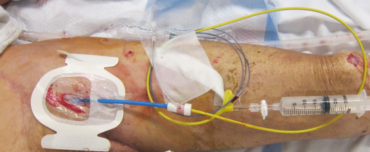
Sounds simple, but there are a number of considerations in this step. The end result is that a guidewire must end up in a large artery somewhere. The common femoral artery (CFA) is the vessel of choice, as anything more distal will rupture during the next step.
There are several ways to access the CFA. In some cases, a femoral arterial line may have already been inserted for other indications. Or maybe “just in case” REBOA might be needed. However, it’s important that the catheter is in the common femoral artery, not the superficial. This means that it must be inserted very close to the inguinal ligament.
If the patient still has vital signs, an arterial line may be inserted quickly, preferably with ultrasound guidance. However, if vitals have been lost, only a cutdown will assure rapid access to the artery.
Step 2. Insert and position the balloon.
First, make sure an x-ray unit is available, and position a plate underneath the patient’s body, from nipples downward. This is helpful for confirming positioning of the guidewire, if used. If not readily available, the wire and balloon can be marked based on external landmarks on the patient’s body.
For external marking, hold the REBOA next to the patient. For Zone I mark the catheter measuring from the groin to the xiphoid. Zone III should be marked with the balloon just above the umbilicus.
Now convert the existing wire to the appropriate size sheath for the REBOA catheter using the manufacturer’s instructions. Insert the REBOA unit, again following the directions for a wired or wireless catheter. Smoothly insert until your catheter mark is at the level of your sheath. Lock everything into place.
Step 3: Inflate the balloon!
Whereas the previous steps only require some degree of technical skill, this one requires good judgment. The key is to get good occlusion without rupturing a vessel (the aorta!). This means inflating until you feel the pressure needed to add more volume start to increase disproportionately. Kind of like adjusting the cuff pressure on an endotracheal tube by feel.
I recommend inflating the balloon, not with plain saline, but saline with a bit of IV contrast mixed in. This allows you to verify balloon position using that x-ray unit and plate you so thoughtfully placed in the last step, or with fluoroscopy in the OR.