All posts by The Trauma Pro

New Trauma MedEd Newsletter Released Soon!

I’m just putting the finishing touches on the next newsletter. It contains everything that you really want to know about Trauma in Pregnancy. Here are the contents:

  • Predicting outcomes
  • Tips & Tricks (for EMS and physicians)
  • Imaging
  • Peri-mortem C-section: when, with what, and how?

I’m going to release this issue to subscribers on Halloween. Everyone else can pick it up here on the blog about 10 days later.

If you want to get it as soon as it is released, please subscribe by clicking here! And you can pick up back issues when you follow the link, too!

tmeimage1116

What You Need To Know About Frontal Sinus Fractures

Fracture of the frontal sinus is less common than other facial injuries, but can be more complex to deal with, both in the shorter and longer terms. These are generally high energy injuries, and facial impact in car crashes is the most common mechanism. Fists generally can’t cause the injury, but blunt objects like baseball bats can.

Here’s the normal anatomy:

sinus-fracture-treatment


Source: www.facialtraumamd.com

There are two “tables”, the anterior and the posterior. The anterior is covered with skin and a small amount of subcutaneous tissue. The posterior table is separated from the brain by the meninges.

Here’s an image of an open fracture involving both tables. Note the underlying pneumocephalus.

frontal_sinus1

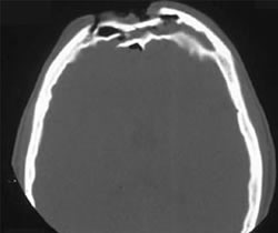
A third of injuries violate the anterior table, and two thirds violate both. Posterior table fractures are very rare. A third of all patients will develop a CSF leak, typically from their nose.

These fractures may be (rarely) identified on physical exam if deformity and flattening is noted over the forehead. Most of the time, these patients undergo imaging for brain injury and the fracture is found incidentally. Once identified, go back and specifically look for a CSF leak. Clear fluid in the nose is, by definition, CSF. Don’t waste time on a beta-2 transferring (see below).

If a laceration is clearly visible over the fracture, or if a CSF leak was identified, notify your maxillofacial specialist immediately. If more than a little pneumocephalus is present, let your neurosurgeon know. Otherwise, your consults can wait until the next morning.

In general, these patients frequently require surgery for the fracture, either to restore cosmetic contours or to avoid mucocele formation. However, these are seldom needed urgently unless the fracture is an open fracture with contamination or there is a significant CSF leak. If in doubt, though, consult your specialist.

Related posts:

Using Your Hybrid OR For Trauma

Every hospital wants some gadget or other. First, it was a robot. Or two. Now, it’s a hybrid operating room.

lourdes-hybrid-or1

What is this, you ask? It’s a mashup of an operating room and an interventional radiology suite. It’s new. It’s big. It’s cool (literally, which is an issue for trauma surgeons).

More and more hospitals are adding hybrid rooms at the request of their vascular surgery teams. These rooms allow for both angiographic and open operative procedures, potentially at the same time. They are perfect for endovascular procedures that need some degree of hands-in work as well. They are frequently used for thoracic endovascular repair of the aorta (TEVAR), repair of abdominal aortic aneurysm (AAA), and transcatheter aortic valve replacement (TAVR).

These rooms would seem to be perfect for some trauma cases as well. Some injuries require a mix of interventional work and open surgery. Think complex pelvic fractures and extremity vascular injuries.

But before you go rushing off to the hybrid room with the next patient you think might benefit from it, consider these issues:

  • You must first secure access to the hybrid room. Just because you want it doesn’t mean you can get it. This room was probably built with other services in mind. You must work with them closely to set up rules and priorities. Consider questions like, can a trauma case bump an elective one?
  • Decide what specific cases can be done in the room. Don’t waste it on procedures that can be done in any old OR. Ideally, it is for multi-team cases and must take advantage of the radiographic capabilities of the hybrid room. If it doesn’t, it should be done in any other room of appropriate size.
  • Check your hardware. Make sure that anything you might attach to the hybrid table actually will attach to it. Frequently, the side rails are missing and the table thickness is different than a standard OR table. Check all of your retractor systems for compatibility. If your neurosurgeons use a skull clamp like a Mayfield, make sure it will attach to the table. If they do not, look for adapters to make it possible. Don’t discover this on your first trip to the room.
  • Watch for hypothermia! These are big rooms, and are difficult to heat up uniformly. In addition, the electronics in the room may be heat sensitive, so you may not be able to raise the temperature to the levels you are accustomed. Place heating systems under and around the patient as much as possible, warm everything that goes into them, and monitor their temp closely.
  • Treat the equipment with respect.  This stuff is delicate, and must be used by other surgeons for sensitive procedures. Don’t break it!

Related posts:

How To Remember Those “Classes of Hemorrhage”

The Advanced Trauma Life Support course lists “classes of hemorrhage”, and various other sources list a similar classification for shock. I’ve not been able to pinpoint where these concepts came from, exactly. But I am sure of one thing: you will be tested on it at some point in your lifetime.

Here’s the table used by the ATLS course:

classes_of_shock

The question you will always be asked is:

What class of hemorrhage (or what % of blood volume loss) is the first to demonstrate systolic hypotension?

This is important because prehospital providers and those in the ED typically rely on systolic blood pressure to figure out if their patient is in trouble.

The answer is Class III, or 30-40%. But how do you remember the damn percentages?

multiscore-maxi1

It’s easy! The numbers are all tennis scores. Here’s how to remember them:

Class I up to 15% Love – 15
Class II 15-30% 15 – 30
Class III 30-40 30 – 40
Class IV >40% Game (almost) over!

Bottom line: Never miss that question again!

Pan Scanning for Elderly Falls?

The last abstract for the Clinical Congress of the American College of Surgeons that I will review deals with doing a so-called “pan-scan” for ground level falls. Apparently, patients at this center have been pan-scanned for years, and they wanted to determine if it was appropriate.

This was a retrospective trauma registry review of 9 years worth of ground level falls. Patients were divided into young (18-54 years) and old (55+ years) groups. They were included in the study if they received a pan-scan.

Here are the factoids:

  • Hospital admission rates (95%) and ICU admission rates (48%) were the same for young and old
  • ISS was a little higher in the older group (9 vs 12)
  • Here are the incidence and type of injuries detected:
Young (n=328) Old (n=257)
TBI 35% 40%
C-spine 2% 2%
Blunt Cereb-vasc inj * 20% 31%
Pneumothorax 14% 15%
Abdominal injury 4% 2%
Mortality * 3% 11%

 * = statistically significant

Bottom line: There is an ongoing argument, still, regarding pan-scan vs selective scanning. The pan-scanners argue that the increased risk (much of which is delayed or intangible) is worth the extra information. This study shows that the authors did not find much difference in injury diagnosis in young vs elderly patients, with the exception of blunt cerebrovascular injury.

Most elderly patients who fall sustain injuries to the head, spine (all of it), extremities and hips. The torso is largely spared, with the exception of ribs. In my opinion, chest CT is only for identification of aortic injury, which just can’t happen from falling over. Or even down stairs. And solid organ injury is also rare in this group.

Although the future risk from radiation in an elderly patient is probably low, the risk from the IV contrast needed to see the aorta or solid organs is significant in this group. And keep in mind the dangers of screening for a low probability diagnosis. You may find something that prompts invasive and potentially more dangerous investigations of something that may never have caused a problem!

I recommend selective scanning of the head and cervical spine (if not clinically clearable), and selective conventional imaging of any other suspicious areas. If additional detail of the thoracic and/or lumbar spine are needed, specific spine CT imaging should be used without contrast.

Related posts:

Reference: Pan-scanning for ground level falls in the elderly: really? ACS Surgical Forum, trauma abstracts, 2016.