All posts by The Trauma Pro

New Technology: Blood Type In 30 Seconds!

This one is really exciting! Blood banks typically keep a significant number of units of O- “universal donor” blood available. These units can be given immediately when a trauma patient in need arrives, since it contains no antigens to the common blood types. It takes anywhere from 5-15 minutes for the blood bank to determine the blood type from the patient’s blood. Then and only then can they begin delivering “type specific” blood that matches the patient’s blood type.

Researchers at the Third Military Medical University in China have developed a paper-based test to determine the ABO type as well as the Rh type (D). Indicators for A, B, and D antigens turn a blue color when they are present, allowing the clinician or blood bank to accurately determine the blood type in 30 seconds. 

Why is this important? O- is an uncommon blood type, with only about 6% of the US population carrying it. Yet blood banks have to keep an inordinate amount in stock “just in case.” Using a blood type test like this could significantly cut down on unnecessary use of this rare O- blood. Unfortunately, it will be 1-2 years before the test is commercially available. I’m sure our nation’s blood bankers can’t wait!

Here’s a brief video that demonstrates how it works.

YouTube player

Reference: A dye-assisted paper-based point-of-care assay for fast and reliable blood grouping. Science Translational Medicine 15 Mar 2017:
Vol. 9, Issue 381, eaaf9209.

How To: Retrograde Urethrogram

I’ve gotten a number of recent requests to repost this easy, DIY guide to the retrograde urethrogram. Enjoy!

One of the hallmarks of urethral injury is blood and the meatus in males. The standard answer to the question “how do you evaluate for it?” is “retrograde urethrogram.” Unfortunately, too few people know how to perform this test, and not all radiologists are familiar. Many times it falls to the urologist, who may not be immediately available.

The technique is simple. This is my variation on the standard technique. The following items are needed:

  • A urine specimen cup
  • A tube of KY jelly (not the little unit dose packs)
  • A bottle of renografin or ultravist contrast
  • A 50-60 cc Toomey syringe (slip-tip)
  • A fluoroscopy suite

Pour 25cc of contrast and 25cc of KY jelly in the specimen cup, cap it and shake well. Draw the contrast jelly up into the syringe. Under fluoro, insert the tip of the syringe into the penis and pull the penis toward yourself, pinching the meatus around the tip of the syringe. Slowly inject all the contrast, watching the contrast column on the fluoro screen. Once there is easy flow into the bladder, you can stop the study. If you see extravasation into the soft tissues, stop the study and call Urology.

The advantages to using this technique are:

  • The contrast/jelly mix creates a contrast gel that is less likely to leak from the meatus when injected
  • The jelly makes it easy to insert the catheter if no urethral injury is detected

Normal urethrogram:

Normal urethrogram

Abnormal urethrogram:

Abnormal urethrogram

Prevnar 13 And Spleen Trauma

Recently, I’ve noticed television commercials for Prevnar-13, a 13-valent pneumococcal vaccine for immunocompromised or asplenic adults. And interestingly, I noticed that the CDC has now added a recommendation such that these patients receive this vaccination, and then the good old 23-valent vaccine (Pneumovax) 8 weeks later.

WTF? Patients with splenectomy (or significant angio-embolization) for trauma are considered functionally asplenic. And although the data for immunization in this group is weak, giving triple vaccinations with pneumcoccal, H. flu, and meningococcal vaccines has become a standard of care.

This was difficult enough already because there was debate around the best time to administer: during the hospital stay or several weeks later after the immune system depression from trauma had resolved. The unfortunate truth is that many trauma patients never come back for followup, and so don’t get any vaccines if they are not given during the hospital stay.

And then came the recommendation a few years ago to give a 5-year booster for the pneumococcal vaccine. I have a hard time remembering when my last tetanus vaccine was to schedule my own booster. How can I expect my trauma patients to remember and come back for their pneumococcal vaccine booster?

So what do we do with the CDC Prevnar-13 recommendation? If we add it, it means that we give Prevnar while the patient is in the hospital, and then hope they come back 8 weeks later for their Pneumovax. And then 5 years later for the booster dose. Huh?

Looking at the package insert, I read that Pneumovax protects against 23 serotypes of S. Pneumo, which represent 85% of most commonly encountered strains out there. So it’s not perfect. Prevnar-13 protects against 13 serotypes, and there is no indication as to what percent of strains encountered are protected against.

So I decided to dig deeper and look at the serotypes included in each vaccine. Here they are:

  • Pneumovax: 1, 2, 3, 4, 5, 6B, 7F, 8, 9N, 9V, 10A, 11A, 12F, 14, 15B, 17F, 18C, 19F, 19A, 20, 22F, 23F, and 33F
  • Prevnar: 1, 3, 4, 5, 6A, 6B, 7F, 9V, 14, 18C, 19A, 19F, and 23F

I bolded the serotypes in Prevnar-13 not found in the Pneumovax vaccine. There was only one, serotype 6A. Unfortunately, it’s nearly impossible to find the prevalence by serotype, and it varies geographically and over time.

Bottom line: I’m not an epidemiologist. But making a set of vaccination rules more complicated for a complex population, and for indications that are a bit weak in the first place, seems unwise. Especially since the added vaccine offers protection for only one more serotype of Pneumococcus.

So please help me out here. Show me something I’m missing. Otherwise, I’ll stick to the original three vaccines, and try to remind my patients to get that booster five years down the road.

Related posts:

Reference: Use of 13-Valent Pneumococcal Conjugate Vaccine and 23-Valent Pneumococcal Polysaccharide Vaccine for Adults with Immunocompromising Conditions: Recommendations of the Advisory Committee on Immunization Practices (ACIP). MMWR 61(40):816-819, October 12, 2012.

Post-Embolization Syndrome In Trauma

A reader requested that I write about post-embolization syndrome. Not being an oncologist or oncologic surgeon, I honestly had never heard about this before, let alone in trauma care. So I figured I would read up and share. And fortunately it was easy; there’s all of one paper about it in the trauma literature.

Post-embolization syndrome is a constellation of symptoms including pain, fever, nausea, and ileus that occurs after angio-embolization of the liver or spleen. There are reports that it is a common occurrence (60-80%) in patients being treated for cancer, and there are a few papers describing it in patients with splenic aneurysm. But only one for trauma.

Children’s Hospital of Boston / Harvard Medical School retrospectively reviewed 12 years of their pediatric  trauma registry data. For every child with a spleen injury who underwent angio-embolization, they matched four others with the same grade of injury who did not. A total of 448 children with blunt splenic injury were identified, and (thankfully) only 11 underwent angio-embolization. Nine had ongoing bleeding despite resuscitation, and two had developed splenic pseudoaneursyms.

Here are the factoids:

  • More of the children who underwent embolization had extravasation seen initially and required more blood products.  They also had longer ICU (3 vs 1 day) and hospital stays (8 vs 5 days). Not surprising, as that is why they had the procedure.
  • 90% of embolized kids had an ileus vs 2% of those not embolized, and they took longer to resume regular diet (5 vs 2 days)
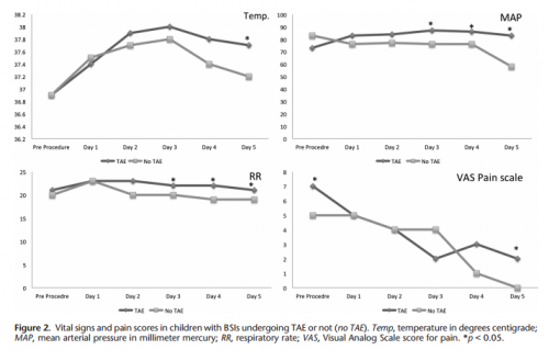
  • Respiratory rate and blood pressure were higher on days 3 and 4 in the embolized group, as was the temperature on day 5 (? see below)
  • Pain was higher on day 5 in the embolized group (? see below again)

Bottom line: Sorry, but I’m not convinced. Yes, I have observed increased pain and temperature elevations in patients who have been embolized. Some have also had an ileus, but it’s difficult to say if that’s from the procedure or other injuries. And this very small series just doesn’t have enough power to convince me of any clinically significant differences in injured children.

Look at the results above. “Significant” differences were only identified on a few select days, but not on the same days across charts. And although the authors may have demonstrated statistical differences, are they clinically relevant? Is a respiratory rate of 22 different from 18? A temp of 37.8 vs 37.2? I don’t think so. And length of stay does not reveal anything because the time in the ICU or hospital is completely dependent on the whims of the surgeon.

I agree that post-embolization syndrome exists in cancer patients. But the findings in trauma patients are too nondescript. They just don’t stand out well enough on their own for me to consider them a real syndrome. As a trauma professional, be aware that your patient probably will experience more pain over the affected organ for a few days, and they will be slow to resume their diet. But other than supportive care and patience, nothing special need be done.

Related posts:

Reference: Transarterial embolization in children with blunt splenic injury
results in postembolization syndrome: A matched
case-control study. J Trauma 73(6):1558-1563, 2012.

Incidental Finding: Gas In The Spleen After Embolization

Most solid organ injury practice guidelines include angioembolization in part of the pathway. But very few require re-imaging at any point to see how the liver or spleen are coming along.

But every once in a while another condition arises, or symptoms worsen unexpectedly, causing us to get another CT scan that includes the abdomen and pelvis. And sometimes we see things that we wouldn’t normally see, like air bubbles in the organ that was embolized.

So what is okay, and what requires some kind of intervention? Our friends at ShockTrauma in Baltimore looked at this in 2001 and can provide some pretty good guidance. They reviewed patients who underwent CT scan both before and after embolization over about 2.5 years. They performed the post-embolization scans for specific indications like fevers, elevated WBC count (!), increasing abdominal pain, or an episode of hypotension. A total of 53 patients were studied.

Here are the factoids:

  • 24 patients underwent embolization of the main splenic artery, 22 had selective embolization of part of the spleen, and 7 had both
  • Splenic infarcts occurred in 63% of patients with main artery embolization, but were large (> 50% of the parenchyma) in only 20% of those
  • Infarcts occurred in 100% of selective embolizations, but were small (< 50%) in 93% of cases
  • Infarcts occurred in 71% of patients with both main and selective embolization, and most were small (80%)
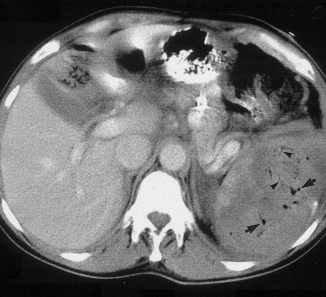
  • Seven (13%) patients developed gas bubbles in the spleen, and was usually present for 1-7 days before disappearing
  • One patient developed increasing gas with pneumoperitoneum and underwent splenectomy for a splenectomy for abscess

This picture that shows tiny bubbles in the spleen parenchyma that represent “normal” gas after embolization:

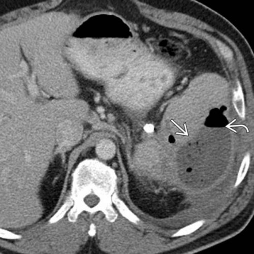
And the following one shows an air/fluid collection in the spleen that indicates an abscess:

Bottom line: Tiny bubbles in the spleen (and probably the liver) occur normally after angioembolization. They usually develop within an area of infarction, and most are benign. It is possible for them to evolve into a splenic abscess, but unlikely. Many embolization patients develop fevers at some point, and most have an elevated WBC count. So in most cases, you can ignore this incidental finding, as long as your patient has mild symptoms.

However, if the patient develops high fevers, very elevated WBC (> 25K), increasing abdominal or flank pain, and the spleen develops an air/fluid level, an abscess is forming. Despite what your radiologist might suggest, catheter drainage is not a good idea. The tubes are too small to remove the slurry that is generally found within the abscess. A trip to the OR is the only effective treatment, and splenectomy is generally the only option.

Related posts:

Reference: CT Findings after Embolization for Blunt Splenic Trauma. J Vasc Intervent Radiol 12(2):209-214, 2001.