All posts by The Trauma Pro

The IVC Filter In Trauma: Why?

The inferior vena cava (IVC) filter has been around in one form or another for over 40 years. One would think that we would have figured everything about it out by now. But no!  The filter has evolved through a number of iterations and form factors over the years. The existing studies, in general, give us piecemeal information on the utility and safety of the device.

One of the major innovations with this technology came with the development of a removable filter. Take a look at the product below. Note the hook at the top and the (relatively) blunt tips of the feet. This allows a metal sheath to be slipped over the filter while in place in the IVC. The legs collapse, and the entire thing can be removed via the internal jugular vein.

ivc-filter-complications1

The availability of the removable filter led the American College of Chest Physicians to recommend their placement in patients with known pulmonary embolism (PE) or proximal deep venous thrombosis (DVT) in patients with contraindications to anticoagulation. Unfortunately, this has been generalized by some trauma professionals over the years to include any trauma patients at high risk for DVT or PE, but who don’t actually have them yet.

One would think that, given the appearance of one of these filters, they would be protective and clots would get caught up in the legs and be unable to travel to the lungs as a PE. Previous studies have taught us that this is not necessarily the case. Plus, the filter can’t stop clots that originate in the upper extremities from becoming an embolism. And there are quite a few papers that have demonstrated the short- and long-term complications, including clot at and below the filter as well as post-phlebitic syndrome in the lower extremities.

A study from Boston University reviewed their own experience retrospectively over a 9 year period. This cohort study looked at patients with and without filters, matching them for age, sex, race, and injury severity. The authors specifically looked at mortality, and used four study periods during the 9 year interval.

Here are the factoids:

  • Over 18,000 patients were admitted during the study period, resulting in 451 with an IVC filter inserted and 1343 matched controls
  • The patients were followed for an average of 4 years after hospitalization
  • Mortality was identical between patients with filters vs the matched controls
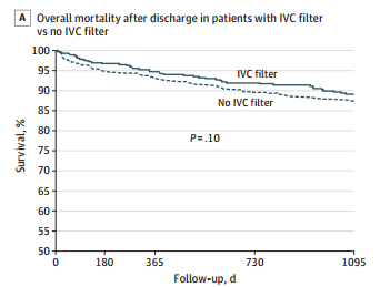
dvt-study

  • There was still no difference in mortality, even if the patients with the filter had DVT or PE present when it was inserted
  • Only 8% ever had their “removable” filter removed (!)

Bottom line: Hopefully, it’s becoming obvious to all that the era of the IVC filter has come and gone. There are many studies that show the downside of placement. And there are several (including this one) that show how forgetful we are about taking them out when no longer needed. And, of course, they are expensive. But the final straw is that they do not seem to protect our patients like we thought (hoped?) they would. It’s time to reconsider those DVT/PE protocols and think really hard about whether we should be inserting IVC filters in trauma patients at all.

Tomorrow: a look at trends in filter insertion and retrieval.

Related post:

Reference: Association Between Inferior Vena Cava Filter Insertion
in Trauma Patients and In-Hospital and Overall Mortality. JAMA Surg, online ahead of print, September 28, 2016.

When Should You Activate Your Backup Trauma Surgeon?

The American College of Surgeons requires all US Trauma Centers to publish a call schedule that includes a backup trauma surgeon. This is important for several reasons:

  • It maintains a high level of care when the on-call surgeon is encumbered with multiple critical patients, or has other on-call responsibilities such as acute care surgery
  • It reduces the need to place the entire trauma center on divert due to surgeon issues

However, the ACS does not provide any guidance regarding the criteria for and logistics of mobilizing the backup surgeon. In my mind, the guiding principle is a simple one:

The backup should be called any time a patient is occupying the on-call surgeon’s time to the extent that they cannot manage the care of a newly arrived (or expected to arrive) patient with critical needs that only the surgeon can provide.

There’s a lot of meat in that sentence, so let’s go over it in detail. 

First, the on-call surgeon must already be busy. This means that they are actively managing one or more patients. Depending on the structure of the call system, they may be involved with trauma patients, general/acute care surgery patients, ICU patients, or a combination thereof. Busy means tied up to the point that they cannot meaningfully manage another patient.

Note that I did not say “evaluate another patient.” Frequently, it is possible to have a resident (at an appropriate training level) or advanced practice provider (APP) see the new patient while the surgeon is tied up, say in the operating room. They can report back, and the surgeon can then weigh his or her choices regarding the level of management that will be needed. Or if operating with a chief resident, it may be possible for the surgeon to briefly leave the OR to see the second patient or quickly check in on the trauma resuscitation. Remember, our emergency medicine colleagues can easily run a trauma activation and provide initial care for major trauma patients. They just can’t operate on them.

What if the surgeon is in the OR? Should they call the backup every time they are doing a case at night? Or every time a trauma activation is called while they are doing one? In my opinion, no. The chance of having a highest level trauma activation called is not that high, and as above, the surgeon, resident, or APP may be able to assess how much attention the new patient is likely to need. But recognize that the surgeon may not meet the 15 minute trauma activation attendance requirement set forth by the ACS.

However, once such a patient does arrive (or there is notification that one of these patients is on the way), call in the backup surgeon. These would include patients that are known to, or are highly suspected of needing immediate operative management. Good examples are penetrating injuries to the torso with hemodynamic problems, or those with known uncontrolled bleeding (e.g. mangled extremity).

If two or more patients are being managed by the surgeon, and they believe that they would not be able to manage another, it’s a good idea to notify the backup that they may be needed. This lets them plan their evening better to ensure rapid availability.

Finally, what is the expected time for the backup to respond and arrive at the hospital to help? There is no firm guideline, but remember, your partner and the patient are asking for your assistance! In my opinion, total time should be no more than 30 minutes. If it takes longer, then the trauma program should look at its backup structure and come up with a way to meet this time frame.

Best of AAST #11: The Need For Trauma Intervention Score (NFTI)

The Trauma Measurement Workgroup at Baylor University in Dallas has been working on a new indicator for identifying major trauma. In a paper published in the Society of Trauma Nursing last year, they determined that six trauma registry variables best identified these patients:

  • transfusion of packed cells within 4 hours of arrival
  • discharge from ED to OR within 90 minutes of arrival
  • discharge from ED to interventional radiology
  • discharge from ED to ICU with a stay > 3 days
  • mechanical ventilation within 3 days, not including OR or procedures
  • death within 60 hours of arrival

Traditionally, Injury Severity Score (ISS) has been used to measure anatomic injury, the Revised Trauma Score (RTS) to quantify physiologic derangement, and their combination (TRISS) to estimate survival.  The authors postulate that physiologic reserve is another determinant of survival, and that NFTI might provide a way to quantify this reserve. One of the QuickShot presentations at the AAST meeting demonstrates how the authors applied this metric.

A multi-institutional data collaborative collected information on more than 88,000 across 35 trauma centers. A complicated mathematical and statistical analysis was carried out, testing how well high ISS (>15), low RTS (<4), and NFTI+ (at least one NFTI variable) predicted mortality, complications, full trauma team activation, length of stay, and procedures performed within 3 days of arrival.

The authors found that NFTI was significantly better at predicting all the outcome variables except full trauma activation than ISS or RTS. And it was still pretty good at that one.

Bottom line: So what does all this mean? The design and analysis of all the numbers is sound. The only thing I take issue with is the assumption that NFTI reflects the “reserve” that a patient has available to combat serious injury. The authors postulate that NFTI is not affected by frailty and comorbidities like ISS and RTS are. I have not seen the manuscript, so perhaps the authors explain the rationale there. But it seems like a stretch. 

What happens if we remove that assumption? Then this study becomes a comparison of a new way to predict resource utilization and/or survival vs ISS and RTS. It uses future variables (as does ISS), so it is difficult to apply this information on patient arrival to treat them any differently, until the first NFTI factor is triggered. But it does predict them well. I think there is considerable potential for NFTI, but we just need more work to make it more useful as early as possible.

Reference: The need for trauma intervention (NFTI) defines major trauma more accurately than injury severity score (ISS) and revised trauma score (RTS): data from a collaboration of 35 adult trauma centers. QuickShot presentation #9, AAST 2018.

Best of AAST #10: Pediatric Contrast Extravasation And Pseudoaneurysms

There is a significant amount of variation in the management of pediatric solid organ injury. This is well documented between adult and pediatric trauma centers in t, but also apparently between centers in different countries. A poster from a Japanese group in Okinawa Japan will be presented this week detailing the relationship between contrast extravasation after spleen or liver injury and pseudoaneurysm formation.

In adults, the general rule is that pseudoaneurysms just about anywhere slowly enlarge and eventually rupture. This group sought to define this relationship in the pediatric age group. They performed a multi-center observational study of retrospectively enrolled children, defined as age 16 and less. Those who had contrast extravasation on initial CT were monitored for later pseudoaneurysm formation.

Here are the factoids:

  • 236 patients were enrolled across 10 participating centers, with about two-thirds having liver injury and the remainder with splenic injury
  • 80% of patients underwent followup CT scan (!!)
  • 33 patients (15%) underwent angiography (!!!!)
  • 17 patients with CT scan (2%) had pseudoaneurysm formation and 4 of them had a delayed rupture
  • Overall, pseudoaneurysms occurred in 29% of those with contrast extravasation and 5% without extravasation
  • The authors concluded that contrast extravasation was significantly associated with pseudoaneurysm formation after adjusting for variables such as ISS, injury grade, and degree of hemoperitoneum

Bottom line: This is an abstract, so a lot is missing. What was the age distribution, especially among those who underwent angiography? Was the data skewed by a predominantly teenage population, whose organs behave more like adults? The abstract answers a question but ignores the clinical significance.

For those trauma professionals who routinely care for pediatric patients, you know that contrast extravasation in children doesn’t act like its adult counterpart. Kids seldom decompensate, and for those who are mistakenly taken for angiography, the extravasation is frequently gone. The authors even admitted in the conclusion that aggressive screening and treatment for pseudoaneurysm was carried out.

The real question is, what is the significance of a solid organ pseudoaneurysm in children? Based on my clinical experience and reading of the US literature, not much. Of course, there is a gray zone as children move into adulthood in the early to mid-teens. But this does not warrant re-scanning and there should be no routine angiography in this age group. Contrast extravasation in pediatric patients warrants close observation for a period of time. But intervention should only be considered in those who behave clinically like they have ongoing bleeding. 

Reference: Association between contrast extravasation on CT scan and pseudoaneurysm in pediatric blunt splenic and hepatic injury: a multi-institutional observational study. Poster 31, AAST 2018.

Best of AAST #9: Popliteal Atery Injury Repair

Injury of the popliteal artery is potentially devastating. Since this vessel is essentially and end artery, any complication resulting in thrombosis can result in limb loss. Traditionally, significant injuries have been treated with open repair and/or bypass. However, endovascular therapies have been making inroads in this area. Short-term outcomes appear to be equivalent. But what happens in the long term? Is one better than the other?

Scripps Mercy in San Diego (yes, same as yesterday’s abstract!) performed a retrospective review of the same California state discharge database. This time, they focused on patients with popliteal artery injury, and the attendant complications of fasciotomy and amputation. They stratified the patients into open and endovascular groups.

Here are the factoids:

  • 769 patients with popliteal artery injury were identified over an 8-year period
  • 59% were managed with an open operation, 4% using endovascular techniques, 2% combined, and 34% nonoperatively
  • Fasciotomies were performed significantly more often in the open group (41% vs 19%)
  • More amputations were performed in open cases, but this was not significantly different (11% vs 3% [1 patient in the endovascular group])
  • Embolism or thrombosis was significantly more likely during the first admission in endovascular or combined endo/open cases
  • Patients requiring both endo and endo+open procedures  were 5x more likely to undergo a later amputation, and 4x more likely to die after discharge

Bottom line: First, remember the limitations of this study: (very) small numbers, and a large database that precludes teasing out details. It suggests that open repair of popliteal injury is superior to endovascular due to higher thrombosis/embolism and amputation rates. Performing a fasciotomy is somewhat subjective, and may be done by surgeon preference to protect the limb. But amputation is more objective.

Unfortunately, we will not get anything more definitive any time soon. This 8-year analysis of a huge state database yielded only 769 cases, or 96 per year. In a state with 39 million people. That’s three injuries (reported) per million people per year. We will never generate a study that will tell us the full answers. But in the meantime, consider endovascular repair of popliteal artery injury only in patients for whom an open procedure is more challenging or risky (e.g. obesity, associated wounds).

Reference: Outcomes for popliteal artery injury repair after discharge: a large-scale population-based analysis. Session XXII Paper 55, AAST 2018.