Confusion At The Trauma Professional’s Blog?

Many readers may have noticed that the blog site has looked different for the past week. The good news is that I’ve migrated all my content (and more) to a standalone website, TheTraumaPro.com.

But the bad news was that all of the search engines only know of the original site, regionstraumapro.com, the original blog hosted on Tumblr. So a lot of people ended up being directed to an old post (on the new site) and not knowing why or how they got there. Confusing! Furthermore, links to related posts on the Tumblr site took readers to the same old random post on the new site. Even more confusing!

In order to stem the confusion while the search engines catch up, I’ve decided to run both sites in parallel. All posts will be cross-posted to both sites simultaneously. The Twitter notification will link to the post on the new site, but it will still be on Tumblr as well.

Please check out all the extra content on the new site at:

TheTraumaPro.com

but just be aware that searches for content will probably direct you to Tumblr at:

regionstraumapro.com

Thanks for reading, whichever one you choose!

Michael

Cervical Spine MRI After Negative CT

dislocation-atlanto-axial-0005

There are multiple ways to clear a cervical spine! Most centers use a combination of clinical decision tools and CT scan in adults. The gold standard tie breaker, warranted or not, seems to be MRI. This tool is only used in select cases where conventional imaging is in doubt, or the clinical exam is puzzling.

Some centers clear based on CT only as long as imaging is indicated. Some use MRI in cases where patients continue to complain of midline neck pain or tenderness after negative CT. A multi-center trial encompassing 8 Level I and II centers prospectively performed MRI on patients who could not be clinically evaluated, or had persistent midline cervical pain after normal CT.

A total of 767 patients were seen over a 30 month period. Besides looking at the usual data points, the authors were interested in new diagnoses and changes in management based on the MRI results.

Here are the factoids:

  • Neck pain and inability to evaluate occurred with equal frequency, about 45%; the remaining 10% had both
  • 23% of MRIs were abnormal, with 17% ligament injury, 4% swelling, 1% disk injury, and 1% dural hematomas.
  • Patients with normal and abnormal MRI had neurologic anomalies about equally (15-19%). [Why are these patients included? Were they initially not evaluable?]
  • The cervical collar was removed in 88% of patients with normal MRI (??), and in 13% with abnormal MRI
  • After (presumably) positive MRI, 14 (2%) underwent spine surgery; 8 of these had neurologic signs or symptoms

Bottom line: I’m a bit confused. If the authors were really trying to figure out the rate of abnormal MRI after negative CT, they should have excluded the patients with known neurologic findings. These patients should nearly always have an abnormal MRI. And why did they not take the collar off of the 12% of patients with both normal CT and MRI??

Hopefully, details in the presentation next week will help explain all this. I suspect that the study will show that there are cases where CT is normal but MRI is not. The abstract does not clearly describe how many of these are clinically significant.

I admit, I’m not very comfortable clearing the cervical spine in a patient with negative CT (even if read by a neuroradiologist) and obvious midline neck pain/tenderness. I hope this study helps clarify this issue. We shall see…

Reference: Cervical spine MRI in patients with negative CT: a prospective, multicenter study of the research consortium of New England centers for trauma (ReCONECT). AAST 2016, Paper 61.

Early Operative Fixation of Pelvic Fractures And Functional Outcome

Disruption of the pelvic bones takes a huge amount of energy, and results in significant bleeding and morbidity from other causes. Repair typically consists of surgical fixation, frequently with temporary external fixation in the interim. These patients require intensive therapy postoperatively, with inpatient rehab prior to discharge home.

How well do patients with severe pelvic fractures do in the longer term? The group at the University of Tennessee in Memphis did a lengthy followup study spanning 18 years of severe pelvic fractures treated at their hospital. These patients had sustained fractures with significant bleeding, an open book component, or SI joint disruption with vertical shear.

open book pelvis pre

The authors used phone interviews and a standardized measurement instrument (Activity Measure for Post-Acute Care, AM-PAC) to gauge daily activity of affected patients. They then looked for factors predictive of functional outcome.

Here are the factoids:

  • 401 patients were identified over the 18 year study period
  • Of these only 71% survived (285), and the study documented followup in 145 (51%)
  • Average ISS was 27 (fairly high) and patients tended to be older (mean 53 years)
  • Even after 8 to 20 years, mobility and activity were significantly impaired as measured by AM-PAC
  • Time to fixation was the only identifiable factor that had an impact on decreased mobility or activity

Bottom line: Early definitive fixation of the pelvis was the only variable found that had an impact on future mobility and activity. Frequently, external fixation is applied soon after admission. But remember, your trauma patient is at their healthiest as they roll through the doors of your ED. The sooner they get all of their problems fixed, the better (and safer).

Impact of early operative pelvic fixation on long-term functional outcome following sever pelvic fracture. AAST 2016, Paper 60.

Spleen Injury, Angiography, And Splenectomy

The shift toward initial nonoperative management of spleen injuries began in the early 1990’s, as the resolution of early CT scans began to improve. Our understanding of the indicators of failure also improved over time, and success rates rose and splenectomy rates fell.

Angiography was adopted as an adjunct to early management, especially when we figured out what contrast extravasation and pseudoaneurysms really meant (bad news, and nearly certain failure in adults). At first, it was used in a shotgun approach in most of the higher grade injuries. But we have refined it over the years, and now it is used far more selectively at most centers.

A group at Indiana University was interested in looking at the impact of angio use on splenic salvage over a long time frame. They queried the National Trauma Data Bank, looking specifically at high grade splenic injury care at Level I and II centers from 2008-2014. Patients undergoing splenectomy were divided into early (<= 6hr after admission) and late (> 6 hrs). Over 50,000 records were analyzed.

Here are the factoids:

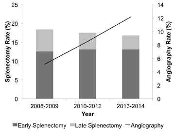
  • There was a shift from early splenectomy to late splenectomy over the study period that was statistically significant
  • Use of angio increased from 5 to 12% during the study period
  • Overall splenectomy rate remained about the same

So the authors recognize that late splenectomy has decreased. But they also state that early splenectomy has increased. They attribute it to increased recognition of patient requiring early splenectomy. They then call into question the need to use angiography if it hasn’t decreased the overall splenectomy rate.

Problem: The early splenectomy rate increased from about 13% to 14%, reading their graph, and is probably not significant. These are the failures that occur in the trauma bay and shortly thereafter that must be taken to the OR. The late splenectomy rate decreased from 5% to 3%, which may be significant (p value not included in the abstract). These are failures during nonoperative management, and are decreasing over time. And BTW, the authors do not define what “high grade” splenic injuries they are looking at.

AAST2016-Paper35

Bottom line: This abstract illustrates why it is important to read the entire article, or in this case, listen to the full presentation at AAST. It sounds like one that’s been written to justify not having angiography available as it is currently required. 

The authors showed that overall splenectomy rate was the same, but delayed splenectomy (late failure) has decreased with increasing use of angiography. But remember, this is an association, not cause and effect. Most of the early failures are still probably ones that can’t be prevented, but we’ll see if the authors can dissect out how many went to OR very early (not eligible for angio), or later in the 6 hour period (could have used angio). It looks to me like the use of angiography is having the desired effect. But undoubtedly we could use that resource more wisely. What we really need are some guidelines as to exactly when a call to the interventional radiologists is warranted.

Related posts:

Reference: Overall splenectomy rates remain the same despite increasing usage of angiography in the management of high grade blunt splenic injury. AAST 2016, paper 35.

Efficacy Of Preperitoneal Packing For Pelvic Fractures

A multi-center trial published in 2015 showed an astounding 32% mortality rate for patients with shock from pelvic fracture. And as I continue to preach, going any place but the OR is dangerous for the patient. Unfortunately, it’s generally not feasible to operatively fix the pelvis acutely, and external fixation has limited impact on ongoing hemorrhage.

If the patient can be stabilized to some degree, interventional radiology can be very helpful. Unfortunately, access after hours involves some degree of time delay. Ideally, the team arrives in 30 minutes or less. But the patient may not be ready, so time to procedure may increase significantly.

So preperitoneal packing of the pelvis (PPP) has now become popular. Years ago, we tried to pack the pelvis from the inside (peritoneal cavity), but it never worked very well. You can push sponges deep into the pelvis as firmly as you want, but the intestines will not keep them from expanding back out of the pelvis.

PPP entails making a lower midline incision, but not entering the peritoneal cavity. A hand is then slid along the anterior surface of the peritoneum around the inside of the iliac wing. Sponges can then be pushed around toward the sacrum, applying direct pressure over bleeding fracture sites and the overlying tissues.

preperitoneal-packing

Image courtesy of ACSSurgery.com

But does it work? Denver Health performed an 11 year retrospective review of their experience with 2293 patients with pelvic fractures. They looked at time to intervention, blood product usage, and mortality.

Here are the factoids:

  • A total of 128 patients underwent PPP
  • Most were younger (mean age 43) and badly injured (mean ISS 48)
  • Median time from door to OR was 44 minutes
  • Patients received an average of 8 units of RBCs intraop, and an additional 3 units in the ensuing 24 hours
  • Overall mortality was 21% (27 of 128), but 9 (7%) were due to severe head injury

Bottom line: Compared to other published studies, time to “definitive management” with PPP was very short. Blood usage also dropped quickly after the procedure. Mortality seems to be much better than expected at about 13%. These results suggest that if you have to wait for angio, or your patient is too unstable to go there, run to the OR first to do some PPP.

And don’t forget these other important management tips:

  • If you see any posterior pelvic fracture on the initial pelvic x-ray, call for blood
  • If the blood pressure softens at any point activate your massive transfusion protocol
  • Apply a binder, especially for open book type fractures
  • Always get a CT in stable patients to help your orthopedic surgeons plan, and to identify contrast blushes
  • If the patient has to go to OR first to stabilize them, consider angio afterwards. You’ll probably find something they can fix.
  • Think about using your hybrid OR!

Reference: Preperitoneal pelvic packing reduces mortality in patients with life-threatening hemorrhage due to unstable pelvic fractures. AAST 2016, Paper 32.