What The Heck? Head CT – The Answer

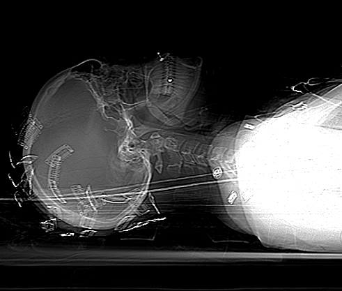
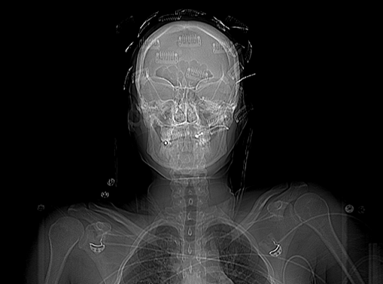
It appears that no one was able to figure this one out! To recap, a young person (female in this case) sustained blunt trauma to the head. When her head was scanned, the following was found on the scout scan:

What is all this odd stuff? They look like some kind of metallic clips placed all over the head. The answer? Hair extensions! Here’s what they look like up close.

Unfortunately, they cannot be left in place during the CT. The amount of scatter is significant enough to degrade the quality of the study. By definition, if you have taken your patient to CT, they are stable and you have a little time. So carefully remove all of the extensions and place them in a bag and save them for the patient (they can be expensive!). 

Make sure the CT tech obtains another topogram to confirm removal of all of the extensions. Then proceed with your CT as usual!

Why Do They Call It: Extra-axial Blood?

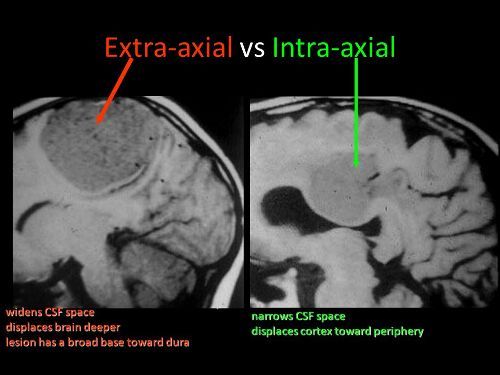
You’ve seen it on head CT reports. “The patient has a collection of extra-axial blood…” Then it goes on to describe the location and size of a subdural hematoma. But why is it called “extra-axial?”

The answer lies in the embryology of the central nervous system. Yes, it’s been a long time since any of us have read anything about that. Early animals had a straight neural tube, which slowly evolved into a brain and spinal cord. This is known as the axis of the nervous system.

The brains of early vertebrates developed at the end of the neural tube, and were oriented in the same longitudinal axis as the rest of it. As brains got bigger, a 90 degree bend occurred at the cephalic flexure.

So in humans, there is a difference between the body axis and the brain axis. But the brain axis is what really counts. This means that any blood outside of the brain axis is defined as extra-axial.

Bottom line: Extra-axial blood is defined as any bleeding outside of the brain parenchyma. This includes subdural and epidural hematomas, and subarachnoid hemorrhage. It does not include any intraparenchymal bleeding like contusions, strokes, or hematomas.

Who Is At Risk For Blunt Cerebrovascular Injury?

Yesterday, I wrote about proper screening for blunt cerebrovascular injury (BCVI). But, as you know, it’s important to screen only when there is a significant risk of the injury being present. Screening using the shotgun approach (screen everyone for everything) yields enough false positive results to present potential danger to your patient.

A variety of authors on this topic have promoted a number of high risk criteria to trigger a screening test. Most make sense, and are related to the anatomy of the vessels in question. The carotid arteries are relatively unprotected, although a bit deep, as they course up the neck. Thus, it is possible to damage them when they suffer a direct and significantly hard blow. Once they enter the skull, they are better protected. However, fractures through key areas of the skull base and face can injure the vessels, even in these protected locations.

The vertebral arteries are deep and relatively protected as they course through the vertebral foramina. However, if the vertebrae are fractured or subluxed, vessel injury can occur.

Finally, and as always, the physical exam is important. If there are unexpected neurologic changes that can’t be explained by other injuries, or there are indications of deep vascular injury, BCVI needs to be considered.

Here is my list of indications to screen for BCVI:

  • Neurologic abnormality not explained by diagnosed injury
  • Arterial epistaxis†
  • Seat belt sign on neck†
  • GCS < 8 (this is the most commonly forgotten one)
  • Petrous bone fracture
  • C‐spine fracture (C1‐C3) or subluxation at any level†
  • Fracture through foramen transversum†
  • LeFort II or III fractures

Bottom line: Be on the lookout for any of the criteria listed above in your trauma patient. If you find one during your initial evaluation, be sure to order a CT angiogram of the neck. And keep an eye out while scanning the head and cervical spine. If any of the other radiographic indications become apparent, add on the CT angiogram at that point.