Practical Tip: Evaluation of Hematuria in Blunt Trauma

Bloody urine is a relatively uncommon finding in blunt trauma patients. Hematuria ranges from microscopic to gross. Microscopic means blood that can only be seen with a microscope, and gross means visible to the naked eye. In trauma, we only care about gross hematuria, which ranges from the faintest of pink to the deepest red.

In the picture above gross hematuria is present in all tubes but the far right one. Those four will need further evaluation.

In trauma, gross hematuria is a result of an injury to kidney, ureter or bladder. Blunt injury to the ureter is so rare it’s reportable, so you can pretty much forget that one unless the mechanism is extreme. So you really just need to focus on kidney and bladder.

Any victim of blunt trauma that presents with visible hematuria needs to be evaluated by CT of the abdomen and pelvis with an added CT cystogram. Standard CT technique is done without a urinary catheter, or with the catheter clamped. This is not acceptable for hematuria evaluation, as only 50% of bladder injuries show up with this technique.

CT cystogram is an add-on to the standard CT, and consists of the administration of contrast into the bladder which is then kept under pressure while the scan is performed. Delayed slices through the pelvis after the bladder is depressurized and emptied is routine. Nearly 100% of bladder injuries are detected using this technique.

If the CT shows a renal laceration or hematoma, the patient should be admitted and managed according to your solid organ injury protocol. Kidney injuries fare better that livers and spleens, and only rarely require surgery. If no kidney or bladder injury is seen, the default diagnosis of a renal contusion is the culprit. No treatment is needed, and the patient can be discharged if no other injuries are present. The blood will clear over a few days, but may disappear and reappear a few times in the process. Be sure to warn the patient that this may occur, or you may receive some surprise phone calls. The patient can followup with their primary care physician in a week or two.

The majority of these injuries do not require urologic consultation. Complex injuries with extravasation of urine out of the kidney, or injuries to the collecting system should be referred to a urologist, however.

Trends In Resident Trauma Operative Experience

Even though it’s called trauma surgery, the operative experience in trauma tends to be somewhat limited. This is due mostly to the fact that most trauma centers see predominantly blunt trauma. Yes, there are hospitals around the world where the penetrating injury load remains high and there is operative experience aplenty.

But in the US, the vast majority of trauma centers see mostly blunt trauma. Surgical residents in the US are required to log 10 operative and 20 nonoperative cases to successfully meet residency completion requirements. And blunt trauma is tending to get less and less operative in nature. A good example is the evolution of blunt solid organ injury to mostly nonoperative management.

So what is happening with surgical resident operative trauma experience? And has there been any impact from the work hour restrictions that have gone into effect in the US? A study from Harborview, Denver Health and Seattle Children’s looked at the ACGME operative logs for surgical residents annually from 1989 to 2010. They combined the data into 5 year blocks, with the last two having work hour restrictions in place.

Some interesting findings:

  • Overall mean caseload of major cases (all types) remained steady at about 925 per resident
  • Mean trauma operative caseload decreased from 76 to 39 (beginning of work hour restrictions)
  • Mean trauma operative caseload remained steady at 39 for the 7 years in which work hour restrictions were in effect
  • The number of intra-abdominal trauma operations decreased from 31 to 17, and the number of liver/spleen operations decreased from 5 and 4 to 3 and 2

Bottom line: Resident trauma operative experience has declined and stabilized in the US. This is due to the evolution of our management of blunt trauma. Unfortunately, this decline will reflect on how well prepared surgeons at outlying hospitals are, and in the quality of emergency surgery they may provide. The impact will be felt most by seriously injured patients who cannot be taken to a high level trauma center initially. We need creative solutions to address this issue, such as mini-clerkships in trauma or structured experiences at high level trauma centers for surgeons in outlying hospitals.

Related post: ED at the busiest hospital in the world!

Reference: ACGME case logs: surgery resident experience in operative trauma for two decades. J Trauma 73(6):1500-1506, 2012.

Stuff You Sterilize Other Stuff With May Not Be Sterile??

When one works in the trauma field, or medicine in general, we deal with the need for sterility all the time. We use equipment and devices that are sterile, and we administer drugs and fluids that are sterile. In surgery, we create sterile fields in which to use this sterile stuff.

In the past few years, we’ve come to the realization that the sterility we take for granted may not always be the case. There have been several cases of contaminated implanted hardware. And most recently, supposedly sterile injectable steroids were found to be contaminated with fungus, leading to several fatal cases of meningitis.

A recent article in the New England Journal of Medicine brings a bizarre problem to light: microbial stowaways in the topical products we use to sterilize things. Most drugs and infused fluids are prepared under sterile conditions. However, due to the antimicrobial activity of topical antiseptics, there is no requirement in the US that they be prepared in this way.

A number of cases of contamination have been reported over the years:

  • Iodophor – contamination with Buckholderia and Pseudomonas occurred during manufacture, leading to dialysis catheter infection and peritonitis
  • Chlorhexidine – contaminated with Serratia, Buckholderia and Ralstonia by end users, leading to wound infections, catheter infections, and death
  • Benzalkonium chloride – contaminated with Buckholderia and Mycobacteria by end users, causing septic arthritis and injection site infections

Bottom line: Nothing is sacred! This problem is scarier than you think, because our most basic assumptions about these products makes it nearly impossible for us to consider them when tracking down infection sources. Furthermore, they are so uncommon that they frequently may go undetected. The one telltale sign is the presence of infection from weird bacteria. If you encounter these bugs, consider this uncommon cause. Regulatory agencies need to get on this and mandate better manufacturing practices for topical antiseptics.

Reference: Microbial stowaways in topical antiseptic products. NEJM 367:2170-2173, Dec 6 2012.

Syncope Workup in Trauma Patients

Syncope accounts for 1-2% of all ED visits, and is a factor in some patients with blunt trauma, especially the elderly. If syncope is suspected, a “syncope workup” is frequently ordered. Just what this consists of is poorly defined. Even less understood is how useful the syncope workup really is.

Researchers at Yale retrospectively looked at their experience doing syncope workups in trauma patients. They were interested in seeing what was typically ordered, if it was clinically useful, and if it impacted length of stay. 

A total of 14% of trauma patients had syncope as a possible contributor to their injury. The investigators found that the following tests were typically ordered in these patients:

  • Carotid ultrasound (96%)
  • 2D Echo (96%)
  • Cardiac enzymes (81%)
  • Cardiology consult (23%)
  • Neurology consult (11%)
  • EEG (7%)
  • MRI (6%)

Most of this testing was normal. About 3% of cardiac enzymes were abnormal, as were 5% of carotid imaging and 4% of echocardiograms. 

Important! Of the patients who underwent an intervention after workup, 69% could have been identified based on history, physical exam, or EKG and did not depend on any of the other diagnostic tests.

Conclusion: Syncope workup is not needed routinely in trauma patients with syncope as a contributing factor. Need for intervention can usually be determined by history, exam and EKG performed in the ED. In this study, $216,000 in excess costs would have been saved!

Reference: Routine or protocol evaluation of trauma patients with suspected syncope is unnecessary. J Trauma 70(2):428-432, 2011.

Pop Quiz – Final Answer

Here’s some background info to go with the xray presented Wednesday:

  • Thin
  • Athlete
  • Epigastric trauma

Put these three together and you get a patient engaged in significant physical activity who was struck in the abdomen. If no pads are involved in the sport, the patient has little padding of their own.

This is a setup for pancreatic or duodenal trauma. This patient presented after being struck in the epigastrium by an elbow during a soccer game. It hurt, but wasn’t bad enough to stop playing. The following day, she was a little sore but felt bloated and started throwing up after breakfast.

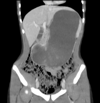
In the ED, a CT was obtained. Here is a coronal view showing the distended stomach:

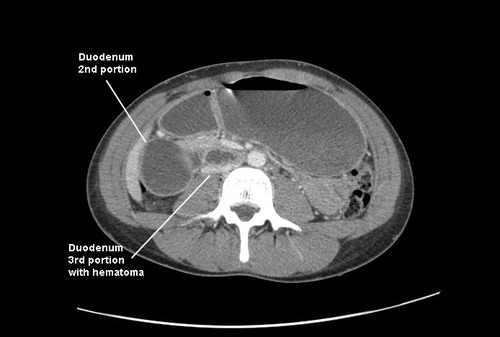
Axial views showed obstruction in the proximal 3rd portion of the duodenum, right over the spine:

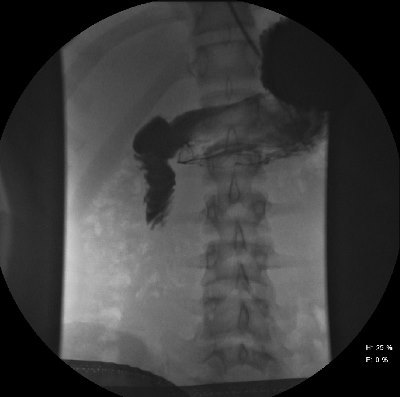
An (unnecessary) contrast study was performed, which confirmed the pathology. Note the tapering and corkscrew appearance of the duodenal folds.


Final diagnosis: duodenal hematoma. This is a crushing injury from compression of the anterior abdominal wall against the spine. The third portion of the duodenum lies over the spine, as does the pancreas, so both are likely to be injured. The latter organ appeared normal on the CT.

Management of blunt duodenal hematoma is simple: wait on it. These will generally resolve quickly over the course of a few days. NG decompression is mandatory, since nothing will pass the obstructed area (saliva, gastic juice, and pancreatic effluent, which add up to 2L+ of fluid per day). In rare cases, parenteral nutrition may be needed if resolution time is approaching the one week mark or in smaller children. A surgical approach with drainage of the hematoma has a low but significant morbidity compared to just waiting. Athletes may return to play soon after recovery.