How Long Should We Watch Intracerebral Hemorrhage?

Patients with traumatic brain injury (TBI) severe enough to cause bleeding are usually admitted to the hospital for observation and in many cases, repeat CT scanning. Those with small intracranial hemorrhages (ICH) may experience progression of the bleeding, and a small percentage of cases may need operative intervention (1-3%). Questions we typically face are, how long should we watch for progression, and how often should we scan?

A retrospective cohort study was carried out at UMD-NJ, looking for answers for a specific subset of these patients. Specifically, they had to have a mild blunt TBI (loss of consciousness and/or retrograde amnesia, GCS in the ED of 13-15) and a positive head CT. They classified any type of hemorrhage into or around the brain as positive. 

During a 3 year period, 474 adults were enrolled but only 341 were eligible for the study. They were excluded due to previous injury, presence of a mass (not trauma), need for immediate neurosurgical intervention, or failure to get a second CT scan. The authors found:

  • 7% of patients were taking anticoagulants! This is surprisingly high. Interestingly, 15 were subtherapeutic, 3 were therapeutic and 2 were supratherapeutic.
  • Subarachnoid hemorrhage was the most common finding on CT (54%). Intraparenchymal hemorrhage was next most common (48%) Many patients had more than one type of bleed.
  • The injury worsened between the first and second scans in 31% of patients. This number increased to 46% in patients taking anticoagulants. 
  • About 97% of bleeds stopped progressing by 24 hrs post-injury.

Bottom line: Most centers are probably overdoing the observation and repeat scan thing. More than two thirds of bleeds are stable by the first scan (first and second scans identical), and nearly all stop progressing within 24 hours. It’s very likely that patients who are not on anticoagulants and who have a stable neuro exam and stable symptoms can get just one scan and 24 hours of observation. Persistent headache, nausea, failure to ambulate well, or other symptoms warrant a repeat scan and longer observation.

Related posts:

Reference: The temporal course of intracranial haemorrhage progression: How long is observation necessary? Injury 43(12):2122-2125, 2012.

Table Of Contents Updated!

Looking for specific content from the Trauma Professional’s Blog? I’ve posted almost 600 items over the last 3 years here, so there’s a lot of stuff to sift through! There are several ways to do it, and here are some tips.

If you look over in the column to the right, you’ll see three tools to help you:

  • The search box. Type in a search term, and Google will do a nice search of just the blog. Note: older versions of Internet Explorer don’t work quite right, so you have to actually click the Search button. Hitting enter will do a Google search of the whole internet, not just the blog.
  • The Indexed archive button. This is a categorized list of all the content on this blog. It’s getting long! I’m in the process of streamling it so it’s more manageable.
  • The Tumblr archive button. This presents a nice thumbnail view of each month’s posts. You can look at all the recent titles, but it’s not really very searchable.
  • The Submit Your Request button. This one is the best! Don’t see what you’re looking for? Send me a request! You’re welcome to email or Skype me as well! I get some of my best material from requests. If you’re curious about a specific topic, other people probably are as well!

Answer: What The Heck Video! Part 2

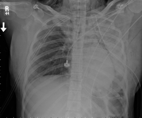
This patient was involved in a motor vehicle crash with significant chest trauma. They’ve been intubated and are oxygenating and ventilating well. What to do next?

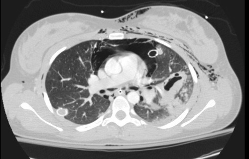
First, the endotracheal tube was a bit deep, which can create its own problems. It was pulled back a few centimeters. Since the patient was hemodynamically stable, a CT angio of the chest would be very helpful to try to figure out the pathology. Here’s a representative slice from the scan.

There are a few striking findings here:

  • Extensive subcutaneous emphysema
  • Large pneummediastinum around the heart
  • Significant injury to the left lung (note the pneumatocele, an air filled collection)
  • Atelectasis of the left lung despite repositioning of the ET tube

The combination of of the above is highly suggestive of a large airway injury. Since the entire lung was affected, it is most likely a mainstem bronchus injury. Usually, these are accompanied by a large air leak from the chest tube, but not in this case.

This prompted the bronchoscopy shown two days ago. The image is oriented such that the left mainstem bronchus was on the right side of the video. A bronchial tear is visible on the lateral aspect, just before the takeoff of the upper lobe bronchus. You can get the impression of a beating heart beating somewhere nearby. And when the camera pops through the laceration, you can actually see the thoracic aortic coursing away toward the diaphragm!

Related post:

Answer: What The Heck Video! Part 1

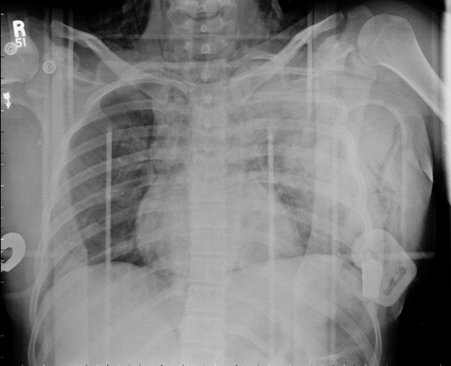
Several of you figured this one out! The patient was involved in a high speed car crash and was brought to the ED in respiratory distress.  Decreased left sided breath sounds were noted. The following xray was obtained.

Opacity is noted in the left lung field, and a modest pneumothorax is seen on the right. Bilateral first ribs are fractured, and the left second through fourth ribs are also broken. The patient was intubated and bilateral chest tubes were inserted.

There was minimal blood from the left chest tube, and a small amount of air from the right. Note the extensive subcutaneous emphysema.

What should we do next? Are any other diagnostic tests indicated? Could the endotracheal tube placement be part of the problem?

Related posts: