This patient was involved in a motor vehicle crash with significant chest trauma. They’ve been intubated and are oxygenating and ventilating well. What to do next?
First, the endotracheal tube was a bit deep, which can create its own problems. It was pulled back a few centimeters. Since the patient was hemodynamically stable, a CT angio of the chest would be very helpful to try to figure out the pathology. Here’s a representative slice from the scan.
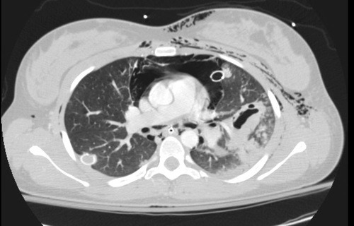
There are a few striking findings here:
- Extensive subcutaneous emphysema
- Large pneummediastinum around the heart
- Significant injury to the left lung (note the pneumatocele, an air filled collection)
- Atelectasis of the left lung despite repositioning of the ET tube
The combination of of the above is highly suggestive of a large airway injury. Since the entire lung was affected, it is most likely a mainstem bronchus injury. Usually, these are accompanied by a large air leak from the chest tube, but not in this case.
This prompted the bronchoscopy shown two days ago. The image is oriented such that the left mainstem bronchus was on the right side of the video. A bronchial tear is visible on the lateral aspect, just before the takeoff of the upper lobe bronchus. You can get the impression of a beating heart beating somewhere nearby. And when the camera pops through the laceration, you can actually see the thoracic aortic coursing away toward the diaphragm!
Related post:
