Tips For Surgeons: Seat Belt Sign

We see seat belt signs at our trauma center with some regularity. There are plenty of papers out there that detail the injuries that occur and the need for a low threshold for surgically exploring these patients. I have not been able to find specific management guidelines, and want to share some tidbits I have learned over the years. Yes, this is based on anecdotal experience, but it’s the best we have right now.

Tips for surgeons:

  • Common injuries involve the terminal ileum, proximal jejunum, and sigmoid colon. My observation is that location in the car is associated with the injury location, probably because of the location of the seat belt buckle. In the US, drivers buckle on the right, and I’ve seen more terminal ileum and buckethandle injuries in this group. Front seat passengers buckle on the left, and I tend to see proximal jejunum and sigmoid injuries more often in them.
  • Seat belt sign on physical exam requires abdominal CT for evaluation, regardless of age. The high incidence of significant injury mandates this test.
  • Seat belt sign plus any anomaly on CT requires evaluation in the OR. The only exception would be a patient with minimal fluid only in the pelvis with an unremarkable abdominal exam. But I would watch them like a hawk.
  • In patients who cannot be examined clinically (e.g. severe TBI), a rising WBC count or lactate beginning on day 2 after adequate resuscitation should prompt a trip to the OR. This is an indirect method for detecting injured bowel or mesentery.
  • Laparoscopy may be used in patients with equivocal findings. Excessive blood, bile tinged fluid, succus, or lots of fibrin deposits on the bowel should prompt conversion to laparotomy. Tip: place all ports distant to the seat belt mark. The soft tissues are frequently disrupted, and gas may leak into this pocket prohibiting good insufflation of the peritoneal cavity.
  • If in doubt, open the abdomen. It’s bad form to put in the scope, see something odd, and walk away. Remember, any abnormal finding after trauma is related to trauma until proven otherwise. It’s almost never pre-existing disease.

Related posts:

Guideline: How To Manage Bleeding In The Anticoagulated Patient

Over the past year, I’ve written about bleeding problems in trauma patients caused or exacerbated by the various anticoagulants now on the market. The field of available drugs keeps growing, and the number of ways to keep blood in liquid form is increasing.

Here’s a link to a set of guidelines for approaching and treating patients who are taking these medications and then develop problematic bleeding. There are few good studies that have actually analyzed the efficacy of these methods, but it’s what we have to work with now. 

If you have any additional maneuvers that you think should be included, please comment or email. And feel free to implement some studies to find the real best practices.

Link: Guideline for bleeding in patients taking anticoagulants

Related posts:

Not Your Usual Pneumothorax?

You’ve been called to the ED to see a patient with a “spontaneous pneumothorax”, but once you meet him you see that he doesn’t fit the classic profile (tall, slim male). What gives?

After closer questioning, he admits to have been smoking crack cocaine at the time. Freak coincidence?

There are a number of case reports dating from 1984 describing this association. A number of reasons have been cited:

  • A high incidence of tobacco smoking
  • Bullous disease caused by inhaled drug use
  • Inhalation of hot gas followed by frequent Valsalva maneuvers

I’ve seen this presentation about 5 times in my career. I always ask about drug use so I can ensure that a chemical dependency screen is ordered.

Reference: Pneumothorax, pneumomediastinum, and pneumopericardium following Valsalva’s maneuver during marijuana smoking. N Y State J Med 84(12):619-20, 1984.

Pediatric Trauma Case: The Answer

So you’ve been called to the ED to see this 10 year old boy who ran into a buddy on the playground while playing tag. They hit chest to chest, but neither had any apparent injuries at the time. Once home, your patient proceeded to cough up a little blood. Mom promptly brought him to your ED for evaluation.

The first thing to do is a good history and physical. No previous illnesses, nothing like this before. No other obvious injuries, no symptoms of concussion. Just some mild anterior chest wall tenderness in the mid-sternum where he hit the other kid.

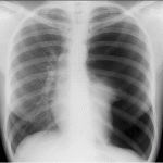
Most likely diagnosis: pulmonary contusion. Now, think about what you need to do and the risks and benefits of the tests you could order. What you need to do is rule out a pneumothorax large enough to be treated. A simple chest X-ray will do this. It won’t detect an occult pneumo, but this is not necessary.

A chest X-ray won’t necessarily show you a pulmonary contusion, either. But do you need to see it to make the diagnosis? No! The clinical evidence is enough. A chest CT is almost never indicated in children, and this is certainly not a reason to get one. EKG: not needed unless your pulse exam was abnormal.

if the child has no complaints of dyspnea and appears to be breathing normally, he can go home. This is such a Low energy injury that progression of the contusion is not an issue. Hospitalization offers no benefit, and will certainly inflict more trauma. Instruct the parents to watch for any apparent breathing problems and give typical non-prescription kiddie analgesics if needed. And be sure to tell them that their son may cough up blood for several more days, but it should disappear soon.

Bottom line: unfortunately, we’ve gotten into the habit of ordering lots of tests to confirm things that we already know. We tend to consider the impact in children a little more, especially when it involves radiation. But we really need to start thinking this way for all patients!