Blood Transfusion With Component Therapy

About 40 years ago, blood banks started moving away from keeping whole blood and began separating it into components (packed cells, platelets, plasma, etc.) for more targeted use. For most uses, this is just fine. But what about trauma?

Trauma patients bleed whole blood. Doesn’t it make sense to give whole blood back? Much of our experience with massive transfusion is derived from our colleagues in the military. Two decades ago, the norm was to give 4 units of packed red cells or so, then give two units of plasma, and every once in a while slip in a bag of platelets. Our military experience seems to indicate that this 4:2:1 ratio is not optimal, and that something like 1:1:1 is better.

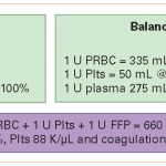
If you think about it, whole blood is already 1:1:1. Splitting it into components and then giving them back seems to be a lot of extra work (and expense) to accomplish the same thing as just giving a unit of whole blood. Plus it triples the exposure to infectious agents and antigens, since the components will usually come from three separate donors. Note that the data in the table above is true for fresh whole blood (not practical in civilian life); banked whole blood will lose some coagulation activity.

Is it time to think about supplying whole blood to trauma centers? And actually looking at whether the outcomes are better or not?

Pneumocephalus And Air Transport

Everybody remembers Boyle’s law, right?

Volume of a gas = k / Pressure     (where K is a constant)

Which means that, as pressure goes down, the volume of a gas increases. This is important for patients who have a pneumothorax and get on an airplane. As the plane ascends the pneumothorax gets bigger and they may have serious problems. Click here to see guidelines on flying after pneumothorax.

Well, what happens if you have air bubbles in your head (pneumocephalus)? Some patients with serious head injury may have this condition but need to be transported by air to definitive care. Most recently, this has been a consideration in military medical evacuation flights out of Afghanistan.

A paper from the US Army and Air Force studied 21 soldiers (small series) who were evacuated by air with known pneumocephalus. The volume of air was estimated by CT prior to transport, and ranged from less than 1ml to 43ml. None of the patients suffered neurologic deterioration during flight, and 3 who had external ventricular drainage (EVD) showed no significant change in intracranial pressure.

Bottom line: Only two cases of tension pneumocephalus have ever been described. Neither occurred in trauma patients. While expanding pneumothorax may be a problem during commercial flight, there is still little data on tension pneumocephalus. It works for the military because the soldiers are in a flying ICU and can be treated immediately if a problem develops. Not so in commercial aircraft, so beware! But remember, medical helicopters don’t fly high enough to create tension problems in any part of the body, so they are not an issue.

Related posts:

Reference: Aeromedical evacuation of patients with pneumocephalus: outcomes in 21 cases. Aviation Space Env Med 79(1):30-35, 2008.

Cricothyroidotomy Using The Scalpel-Bougie Technique

Here’s a video from our colleagues in Australia that shows a slick way of performing a surgical cricothyroidotomy. The number of required instruments is the bare minimum: a scalpel and a bougie. I have not tried this technique, but it looks like it would be very handy when dealing with obese patients with a deep neck. It would also be useful to prehospital providers who are credentialed for crichs and are faced with a difficult airway.

If any of you have used this technique, please leave a comment for us!

Damage Control Dressing: The ABThera (Video)

In the late 1980’s, when we started the work that would be published in the first damage control paper from Penn, we used the vacuum pack dressing. This was first described in a paper from the University of Tennessee at Chattanooga in 1995. Prior to that, the so-called Bogota bag was the usual technique. This consisted of slicing opening up a sterile IV bag (either the standard 1 liter or the urology 3 liter bag for big jobs) and sewing it into the wound. This worked, but it freaked out the nurses, who could see the intestines through the print on the clear plastic bag.

The vacuum pack was patient friendly, with a layer of plastic on the bottom, some absorbent towels in the middle with a drain in place to remove fluid and apply suction, and an adherent plastic layer on top to keep the bed clean. As you can imagine, this was a little complicated to apply correctly. One misstep and things stuck to the bowel or leaked out onto the bed.

In the past few years, a commercial product was developed that incorporated all these principles and was easy to apply. This is the KCI ABThera (note: I have no financial interest in KCI or this product; I just wish I had invented it). The only downside is that there is a small learning curve when first using this product.

YouTube player

The video above shows a demonstration of the application on an abdominal mannikin. It is not as slick as the company videos, but I think it’s more practical, with some good tips.

References:

  • Damage control: an approach for improved survival in exsanguinating penetrating abdominal injury. J Trauma 35(3):375-382, 1993.
  • Temporary closure of open abdominal wounds. Am Surg 61(1):30-35, 1995.