Pulmonary Edema After Chest Tube Insertion

Re-expansion pulmonary edema is an uncommon event after chest tube insertion. Typically, patients have had symptoms of pneumothorax for several days, usually 3 or more. It occurs most often if a large amount of air (or blood) is evacuated at once. The patient will typically become symptomatic within an hour, with decreased oxygen saturation and subjective breathing difficulty.

Although the mechanism is not entirely clear, it appears that the small blood vessels in the lung become more permeable if they are collapsed for an extended period. Mechanical stress from rapid re-expansion further damages the vessels, allowing them to leak. This leads to oxygenation and ventilation problems if severe. 

Practical tips:

  • Check the history. Most of these patients have had their pneumothorax for 3 or more days.
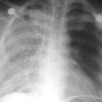
  • Check the xray. Complete pneumothorax (or large hemothorax) puts the patient at high risk.
  • Modify your chest tube insertion technique. Clamp the distal end of the tube so the pneumothorax is not evacuated suddenly as the tube goes in.
  • Modify the collection system. Do not use suction initially; only set up for water seal. Clamp the tubing on the patient side. Every 10-15 minutes release the clamp and briefly let some of the air out of the chest, then reclamp. Repeat this until all air has bubbled through the water seal chamber. 
  • Watch your patient. If they cough excessively, start to desaturate or become dyspneic, get your respiratory adjuncts. Give higher inspired oxygen by appropriate means, and consider BiPap or CPAP. In extreme cases intubation may be needed. If the patient does not have any difficulties after about an hour, connect the collection system to suction and proceed as you normally would.

Reference: Reexpansion pulmonary edema. Ann Thoracic Cardiovasc Surg 14:205-209, 2008.

Trauma Patient Stability

EMS in the field and physicians in the ED are faced with rapidly assigning some degree of stability to the patients they treat. What exactly are the shades of stability, and what considerations are there for each degree?

In my mind, there are three levels of “stability”:

  • Unstable – this one is easy to figure out. The patient has obvious physiologic compromise, which may be objective (low blood pressure, low GCS or poor neuro exam, etc) or subjective (just plain looks bad). 
    EMS: These patients need transport to an appropriate level trauma center (I or II) immediately. If they need airway control or IV access that can’t be obtained in the field, stop at the nearest Level III or IV for assist, then continue on your way FAST. 
    ED: These patient must be a trauma activation. If not activated as your top-tier trauma, activate or upgrade now! These patients must be seen by a trauma surgeon immediately, and can only go to the OR. No diagnostics outside the resuscitation room are allowed unless they can be converted into one of the two stability levels below.
  • Stable – this one is usually easy to figure out, too. These patients look good, have good vitals, and a low to moderate energy mechanism for their trauma. Look out for those few patients that may be hiding something like moderate bleeding into some body cavity.
    EMS: Follow your usual transport protocols to select the closest, appropriate hospital.
    ED: Follow your standard protocols for trauma activation if needed. Transport for standard imaging is fine.
  •  Metastable – this is a term I invented. It describes patients who have evidence of ongoing volume loss that can be controlled with infusion of crystalloid and/or blood products. It is possible to maintain a certainly level of stability using higher than normal volume infusions. This allows physicians to consider diagnostics or interventions outside of an OR.
    EMS: Ensure adequate IV access and give fluids and/or blood per your local protocols. Transport to a Level I or II trauma center as quickly as possible.
    ED: Activate or upgrade to your highest level of trauma activation. The trauma surgeon needs to be present to help direct diagnostics or interventions. These patients may go to CT, IR or other appropriate areas with nurse and physician accompaniment to diagnose and possibly treat bleeding. If the patient changes to unstable at any point, they must immediately be taken to the OR.

I am interested in other opinions on this as well. Please post your comments!

From the Archives: Cervical Spine Imaging

So far, I’ve posted more than 200 items during the past year and a half. There’s a lot of good stuff in the archives, and I am going to periodically provide a list of links to them so they are not forgotten. To see a full index of the archive by subject, click here.

Today, I’m going to focus on cervical spine imaging. Here are four interesting posts from the archives:

As always, I welcome suggestions for new posts!

Using Mechanism of Injury In Your Trauma Activation Criteria

The Centers for Disease Control and Prevention (CDC) published a set of Guidelines for Field Triage two years ago. Click here to download them. They list 4 tiers of activation criteria to help prehospital providers triage patients appropriately to trauma centers. 

Tier 1, which are physiologic criteria, and Tier 2 (anatomic criteria) are very accurate in predicting injury serious enough to require trauma team activation. Tier 3 contains mechanism criteria, and many centers who use these verbatim in their activation criteria end up with a fair amount of overtriage. Some centers even see a significant number of patients who meet Tier 3 criteria go home from the ED!

The Yale department of Emergency Medicine looked at intrusion into vehicle criteria (more than 12" near an occupant, more than 18" anywhere on the vehicle) to see if they are a valid predictor for admission or trauma center transport. It was a retrospective review of EMS transports to the Yale ED or to one satellite site. 

Unfortunately, the number of vehicles that met intrusion criteria (48) was small compared to the number without significant intrusion (560). This makes the data a little less convincing than it may have been. The likelihood that intrusion would require trauma center admission (Positive Predictive Value) was only 26%. The likelihood that trauma center resources would be utilized (for issues like death, ICU stay, operation, spinal injury or intracranial hemorrhage) was only 13%. The authors recommend that the CDC guidelines be tweaked based on this data.

Bottom line: I think the numbers are far too small to convince the CDC to change their guidelines. But I would urge each trauma center that uses the intrusion criteria for activation to carefully study how many of those patients have minor injuries or go home from the emergency department. They may find that they can rely on other more accurate criteria and decrease their overtriage rate at the same time.

Reference: Motor vehicle intrusion alone does not predict trauma center admission or use of trauma center resources. Prehospital Emerg Care 15:203-207, 2011.

EAST Guidelines Update: Spleen Injury

The Eastern Association for the Surgery of Trauma is in the process of updating their trauma practice guidelines for spleen injury. The first set of guidelines was introduced in 2003, and several advances in management have occurred since. here is a summary of the current status of the guidelines:

Level I recommendations (best quality data): 

  • none

Level II recommendations (good data):

  • Initial management of hemodynamically stable patients should be nonoperative
  • Unstable patients should undergo immediate operation or angiographic embolization (my interpretation: unstable patients belong in the OR, not the angio suite!)
  • Patients with peritonitis should go to the operating room
  • Age, grade of injury, amount of hemoperitoneum and age are not contraindications to nonoperative management. Only hemodynamic stability matters.
  • CT of the abdomen with IV contrast is the most reliable method to assess severity of spleen injury (my interpretation: in the hemodynamically stable patient)
  • Angiography with embolization should be considered if a contrast blush is seen on CT, AAST grade > 3, moderate hemoperitoneum is present, or there is evidence of ongoing bleeding
  • Nonoperative management should only be considered if continuous monitoring and serial exams can be carried out at your hospital, and if an operating room is immediately available if needed

Level III recommendations (weak data):

  • Clinical status should dictate need and frequency of followup imaging (my interpretation: only do it if the patient condition changes for the worse)
  • Contrast blush is not an absolute indication for operation or angio-embolization. Age, grade of injury and presence of hypotension need to be considered. (My interpretation: don’t operate or do angio on kids without a really good reason)
  • Angio is an adjunct to nonop management in patients who are at high risk for delayed bleeding or to look for vascular injuries (pseudoaneurysms) that may lead to rupture or delayed hemorrhage

Reference: Trauma Practice Guideline Update, 24th Annual Scientic Assembly, Eastern Association for the Surgery of Trauma, January 2011.